. 2006 Jan;5(1):29-38.
doi: 10.1158/1535-7163.MCT-05-0126.

Inhibition of TRIP1/S8/hSug1, a component of the human 19S proteasome, enhances mitotic apoptosis induced by spindle poisons

Affiliations

Inhibition of TRIP1/S8/hSug1, a component of the human 19S proteasome, enhances mitotic apoptosis induced by spindle poisons

Hiroshi Y Yamada et al. Mol Cancer Ther. 2006 Jan.

Abstract

Mitotic spindle poisons (e.g., Taxol and vinblastine), used as chemotherapy drugs, inhibit mitotic spindle function, activate the mitotic spindle checkpoint, arrest cells in mitosis, and then cause cell death by mechanisms that are poorly understood. By expression cloning, we identified a truncated version of human TRIP1 (also known as S8, hSug1), an AAA (ATPases associated with diverse cellular activities) family ATPase subunit of the 19S proteasome regulatory complex, as an enhancer of spindle poison-mediated apoptosis. Stable expression of the truncated TRIP1/S8/hSug1 in HeLa cells [OP-TRIP1(88-406)] resulted in a decrease of measurable cellular proteasome activity, indicating that OP-TRIP1(88-406) had a dominant-negative effect on proteasome function. OP-TRIP1(88-406) revealed an increased apoptotic response after treatment with spindle poisons or with proteasome inhibitors. The increased apoptosis coincided with a significant decrease in expression of BubR1, a kinase required for activation and maintenance of the mitotic spindle checkpoint in response to treatment with spindle poisons. Small interfering RNA (siRNA)-mediated knockdown of TRIP1/S8/hSug1 resulted in a reduction of general proteasome activity and an increase in mitotic index. The siRNA treatment also caused increased cell death after spindle poison treatment. These results indicate that inhibition of TRIP1/S8/hSug1 function by expression of a truncated version of the protein or by siRNA-mediated suppression enhances cell death in response to spindle poison treatment. Current proteasome inhibitor drugs in trial as anticancer agents target elements of the 20S catalytic subcomplex. Our results suggest that targeting the ATPase subunits in 19S regulatory complex in the proteasome may enhance the antitumor effects of spindle poisons.


Figures

Fig. 1
Fig. 1
Expression of truncated TRIP1/S8 sensitizes cells to spindle poison (A) Schematic presentation of TRIP1/S8/hSug1 and the pSC3 product TRIP1(88-406) that lacks 87 amino acids at the N terminus. TRIP1/S8 is an AAA (ATPases Associated with a variety of cellular Activities) family ATPase subunit found in the 19S regulatory complex of the proteasome. (B) Stable expression of a fragment of the TRIP1/S8 proteasome protein increases cell sensitivity to nocodazole and Taxol in a colony formation assay. The OP-TRIP1(88-406) line, which stably expresses the fragment of the TRIP1/S8 protein (Supplementary figure 1), shows decreased cell proliferation compared to the control cells when challenged with nocodazole or Taxol at low concentration for eight days. Representative plates are shown in left. Cell proliferation was quantified and presented as percentages normalized to zero drug dose. Black bars represent controls. Grey bars represent the OP-TRIP1(88-406) cell line. It is likely that the truncated form of TRIP1/S8 acts in a dominant negative fashion since overproduction of the full length TRIP1/S8 did not significantly affect spindle poison sensitivity (see Supplementary figure 2).
Fig. 2
Fig. 2
Stable expression of an N-terminal truncation of the proteasome subunit TRIP1/S8 results in decreased proteasome activity and elevated sensitivity to proteasome inhibitors. (A) Proteasome activity is reduced in OP-TRIP1(88-406) cells in both asynchronous culture and in cells treated with nocodazole. Cell extracts were prepared and incubated with fluorogenic proteasome substrate III (SucLLVY-AMC) for 30 and 60 minutes in 37°C, and fluorescence was measured. The conversion rate was calculated and proteasome activity in extracts from OP-TRIP1(88-406) cells was compared with extracts from control HeLa cells in both normal cultures (left panel) and in cultures treated with nocodazole (100ng/ml, 16 hours) (right panel). (B) OP-TRIP1(88-406) cells show markedly decreased cell proliferation compared to the parental line when treated with the proteasome inhibitor MG132 (upper panel) or ALLN (lower panel) for eight days. Experiments were repeated at least three times and a typical result is shown. Quantification of cell proliferation is shown in the right panel. Cell proliferation was normalized to zero drug dose. Black bars: Control Hela cells. Grey bars: OP-TRIP1(88-406) cells.
Fig. 3
Fig. 3
OP-TRIP1(88-406) cells undergo accelerated mitotic apoptosis in the presence of spindle poisons. (A) OP-TRIP1(88-406) cells show increased apoptosis when examined after treatment with nocodazole. HeLa and OP-TRIP1(88-406) cells were synchronized in early S phase with a double aphidicolin block, and released into medium containing nocodazole (100ng/ml). Cell morphology was monitored by phase contrast microscopy at the indicated time points. Rounded cells with smooth surfaces were scored as normal mitotic cells. Cells with membrane protrusions (blebs) were scored as apoptotic (dark grey). (B) An example of a OP-TRIP1(88-406) cells undergoing apoptosis. Cells were synchronized and released into nocodazole as in (A), and filmed with time-lapse video microscopy. Video recording was initiated 3 hours after the cells under observation had entered mitosis in the presence of nocodazole (time 3:00). Most OP-TRIP1(88-406) cells arrested in mitosis for 3.5-5.8 hours and then initiated membrane blebbing. Most parental HeLa cells remained arrested in mitosis without showing blebbing for at least 10 hours (not shown). Magnification bar 20μm. (C) Apoptosis was initiated during nocodazole-mediated mitotic arrest. HeLa and OP-TRIP1(88-406) cells were treated with 100ng/ml nocodazole for 4 hours. Rounded mitotic cells were collected and further cultured in medium containing nocodazole (100ng/ml). At the indicated times, cell extracts were prepared, proteins were separated by SDS-PAGE and analyzed by immunoblotting. Equal amounts of protein were loaded in each lane. OP-TRIP1(88-406) cells show accelerated cleavage of PARP, dephosphorylation and loss of the Cdc27 protein, and loss of BubR1 protein while Cyclin B levels remain high during continued incubation in nocodazole. (D) Comparison of FACS profiles revealed an increase in PARP fragment-positive cells with G2/M DNA content in nocodazole-treated OP-TRIP1(88-406) cells compared to controls. We treated control and OP-TRIP1(88-406) cells without or with nocodazole (100ng/ml) for 16 or 24 hours and collected samples for FACS analysis. The DNA content of all sample cells is displayed in upper row (All cells). The samples are also labeled with anti-PARP fragment antibody then by FITC secondary antibody. In some panels, percentage of cells with G2/M DNA content is shown as inset.
Fig. 4
Fig. 4
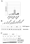
Treatment of HeLa cells with spindle poisons and a proteasome inhibitor increases cytotoxicity and apoptosis. (A) MG132 enhances cell death due to spindle poisons. HeLa cells were treated with nocodazole (100ng/ml), MG132 (10μM) or, Taxol (20nM) alone or in combination with MG132 (10μM) for 16 hours. Cell death was scored using fluorescent markers of membrane permeability (Molecular probes Live/Dead cell death assay kit). (B) Immunoblot for an apoptosis marker, PARP fragment. HeLa cells were treated with nocodazole (100ng/ml; noc100, or 50ng/ml, noc50) alone, MG132 (1μM; MG1) alone, Taxol (20nM; Tax20, or 10nM; Tax10) alone, nocodazole and MG132 (noc100+MG1, or noc50+MG1), or Taxol and MG132 (Tax20+MG1, or Tax10+MG1) simultaneously for 16 hours, and monitored for generation of PARP fragment. β-tubulin is shown as a loading control. (C) Inactivation of the proteasome after mitotic arrest in HeLa cells does not significantly increase the level of apoptosis as monitored by PARP fragmentation. HeLa cells were treated with nocodazole (100ng/ml) or Taxol (1 μM) for 4 hours and mitotic cells were collected by shake off. The mitotic cells were then incubated in the same microtubule drug for an additional 4 and 8 hours with or without the addition of MG132 (10μM). Extracts were prepared and blotted for PARP fragmentation.
Fig. 5
Fig. 5
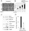
SiRNA-mediated TRIP1/S8 repression results in mitotic cell accumulation, enhanced cell killing with spindle poison treatment, and compromised proteasome function (A) Cellular phenotype. HeLa cells were transfected with siRNA against GFP (Control) or against TRIP1/S8 (TRIP1). Forty hours later cells were incubated with or without 100ng/ml nocodazole, cultured further for eight hours, and observed by phase contrast microscopy. Mitotic cells have smooth round morphology. Note cells in lower right panel (TRIP1 siRNA, nocodazole+) show many apoptotic cells with membrane blebbing. Magnification bar is 20μm. (B) Phenotype quantification. Cells treated as in (A) were stained with fluorescent Annexin V and propidium iodide (PI), and categorized into three phenotypes: Annexin V positive (black area: early apoptotic cells), Mitotic (grey area: rounded, healthy mitotic cells) and Necrotic (PI-positive; textured area: Necrotic cells and apoptotic cells in terminal stages). The sum of three categories represents the total rounded cells. (C) Immunoblots. Cell extracts were prepared from the treated cultures and monitored for TRIP1/S8, PARP fragment (apoptosis marker), Cdc27, phosphorylated histone H3 (mitotic marker) and β-tubulin (loading control). Ct:Control, Tr:TRIP1. (D) In TRIP1/S8 siRNA-treated cells proteasome activity was reduced by 60% compared with control. HeLa cells were transfected with siRNA against GFP (negative control, black bars) or against TRIP1/S8 (grey bars). Forty eight hours after transfection, cell extracts were prepared and, after equalizing the protein amount, were incubated with fluorogenic proteasome substrate III (SucLLVY-AMC) at 37°C for 30 and 60 minutes. The conversion rate was calculated and normalized to control transfectants.

References

    1. Bhalla KN. Microtubule-targeted anticancer agents and apoptosis. Oncogene. 2003;22:9075–86. - PubMed
    1. Jordan MA, Wilson L. Microtubules as a target for anticancer drugs. Nat Rev Cancer. 2004;4:253–65. - PubMed
    1. Mollinedo F, Gajate C. Microtubules, microtubule-interfering agents and apoptosis. Apoptosis. 2003;8:413–50. - PubMed
    1. Blagosklonny MV, Fojo T. Molecular effects of paclitaxel: myths and reality (a critical review) Int J Cancer. 1999;83(2):151–6. - PubMed
    1. Bharadwaj R, Yu H. The spindle checkpoint, aneuploidy, and cancer. Oncogene. 2004;23:2016–27. - PubMed

Publication types

MeSH terms