

Genetic polymorphism and protein conformational plasticity in the calmodulin superfamily: two ways to promote multifunctionality
- PMID: 16432210
- PMCID: PMC1360552
- DOI: 10.1073/pnas.0508640103
Genetic polymorphism and protein conformational plasticity in the calmodulin superfamily: two ways to promote multifunctionality
Abstract
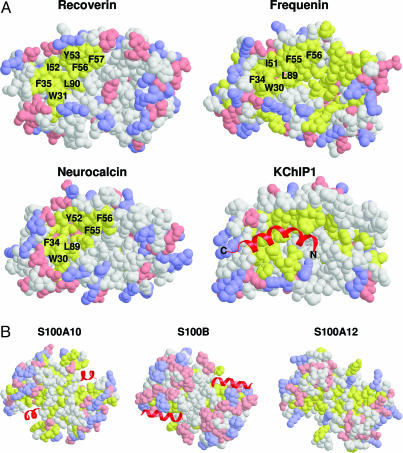
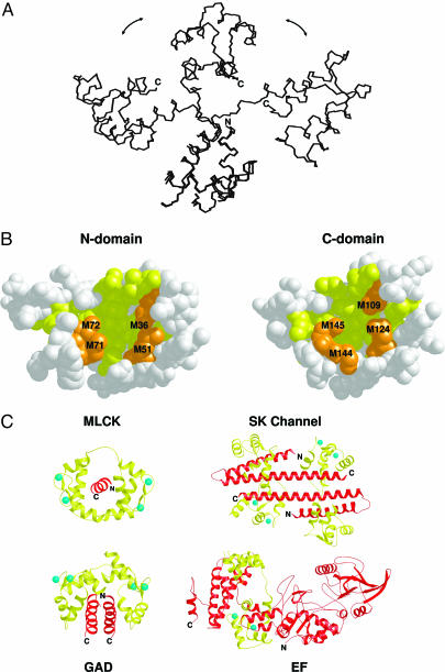
Calcium signaling pathways control a variety of cellular events such as gene transcription, protein phosphorylation, nucleotide metabolism, and ion transport. These pathways often involve a large number of calcium-binding proteins collectively known as the calmodulin or EF-hand protein superfamily. Many EF-hand proteins undergo a large conformational change upon binding to Ca(2+) and target proteins. All members of the superfamily share marked sequence homology and similar structural features required to sense Ca(2+). Despite such structural similarities, the functional diversity of EF-hand calcium-binding proteins is extraordinary. Calmodulin itself can bind >300 different proteins, and the many members of the neuronal calcium sensor and S100 protein families collectively recognize a largely different set of target proteins. Recent biochemical and structural studies of many different EF-hand proteins highlight remarkable similarities and variations in conformational responses to the common ligand Ca(2+) and their respective cellular targets. In this review, we examine the essence of molecular recognition activities and the mechanisms by which calmodulin superfamily proteins control a wide variety of Ca(2+) signaling processes.
Figures
References
-
- Carafoli, E. (2005) FEBS J. 272, 1073–1089. - PubMed
-
- Bootman, M. D. & Berridge, M. J. (1995) Cell 83, 675–678. - PubMed
-
- Berridge, M. J., Bootman, M. D. & Roderick, H. L. (2003) Nat. Rev. Mol. Cell Biol. 4, 517–529. - PubMed
-
- Berridge, M. J., Lipp, P. & Bootman, M. D. (2000) Nat. Rev. Mol. Cell Biol. 1, 11–21. - PubMed
-
- Haiech, J., Moulhaye, S. B. & Kilhoffer, M. C. (2004) Biochim. Biophys. Acta 1742, 179–183. - PubMed
Publication types
MeSH terms
Substances
LinkOut - more resources
Full Text Sources
Other Literature Sources
Miscellaneous
