. 2006 Jan 31;103(5):1382-7.
doi: 10.1073/pnas.0506911103. Epub 2006 Jan 23.

Active digestion of sperm mitochondrial DNA in single living sperm revealed by optical tweezers

Affiliations

Active digestion of sperm mitochondrial DNA in single living sperm revealed by optical tweezers

Yoshiki Nishimura et al. Proc Natl Acad Sci U S A. .

Abstract

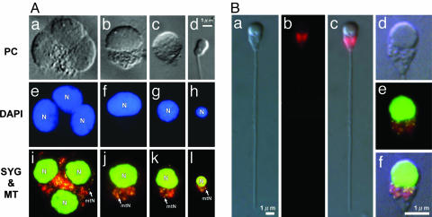
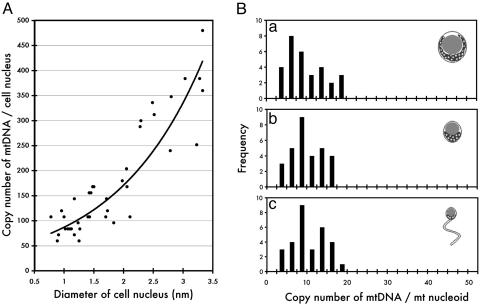
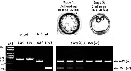
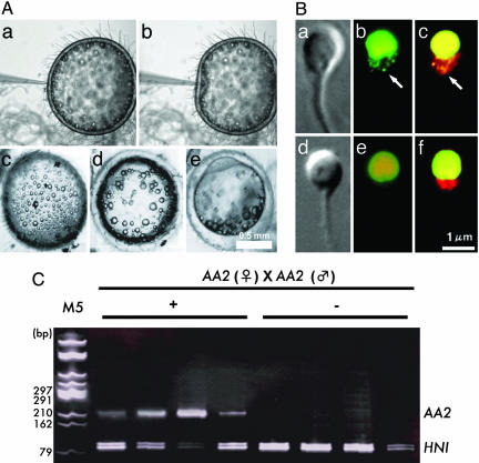
In almost all eukaryotes, mitochondrial (mt) genes are transmitted to progeny mainly from the maternal parent. The most popular explanation for this phenomenon is simple dilution of paternal mtDNA, because the paternal gametes (sperm) are much smaller than maternal gametes (egg) and contribute a limited amount of mitochondria to the progeny. Recently, this simple explanation has been challenged in several reports that describe the active digestion of sperm mtDNA, down-regulation of mtDNA replication in sperm, and proteolysis of mitochondria triggered by ubiquitination. In this investigation, we visualized mt nucleoids in living sperm by using highly sensitive SYBR green I vital staining. The ability to visualize mt nucleoids allowed us to clarify that the elimination of sperm mtDNA upon fertilization is achieved through two steps: (i) gradual decrease of mt nucleoid numbers during spermatogenesis and (ii) rapid digestion of sperm mtDNA just after fertilization. One notable point is that the digestion of mtDNA is achieved before the complete destruction of mitochondrial structures, which may be necessary to avoid the diffusion and transmission of potentially deleterious sperm mtDNA to the progeny.


Figures

Fig. 1.
Fig. 1.
mt nucleoids during spermatogenesis. (A) Phase-contrast (PC) (a–d), DAPI (e–h), and SYBR green I/MitoTracker CMTMRos double-stained (i–l) spermatid cells (a–c, e–g, and i–k) at various developmental stages and matured sperm (d, h, and l). DAPI staining allows visualization of only the cell nucleus (N), whereas SYBR green I staining allows visualization of minute yellow spots (≈1 μm) outside of the cell nucleus. The yellow spots precisely overlap with the red fluorescence of MitoTracker CMTMRos at every developmental stage, indicating that they are mt nucleoids (mtN). (B) Phase-contrast (a) and MitoTracker CMTMRos-stained (b) images of matured sperm. (c) An overlay of a and b.(d and e) Magnified phase-contrast (d) SYBR green I/MitoTracker CMTMRos double-stained (e) images of the head part of matured sperm. (f) An overlay of d and e.
Fig. 2.
Fig. 2.
Fluorescence intensity of mt nucleoids measured by a video-intensified microscope photon-counting system (VIMPCS). (A) Copy number of mtDNA measured by VIMPCS (61) was plotted against the diameter of the cell nucleus. A least-squares fit line calculated by Microsoft excel shows a rapid (≈8- to 10-fold) reduction of the mtDNA copy number along with the condensation of the cell nucleus during the development of sperm cells. (B) Histograms show the distribution of fluorescence intensity of mt nucleoids. The spermatid cells were arbitrarily classified into three groups: before polarization (a), after polarization (b), and matured sperm (c), and the distribution of fluorescence intensity of mt nucleoids was analyzed. Despite the dramatic reorganization of the cell nucleus and the rapid reduction in the number of mt nucleoids per cell along with the development, the fluorescence intensity of mt nucleoids remained almost unchanged.
Fig. 3.
Fig. 3.
Polymorphism in NADH dehydrogenase sequence between AA2 and HNI. (Lower Left) Two hundred and forty-nine bp of NADH dehydrogenase sequence was amplified from AA2 and HNI by nested PCR (uncut) and digested by HinfI (Hinf I cut). Because of the polymorphism, AA2 gave 222- + 27-bp fragments (the latter are too small to be detected), and HNI gave 129- + 120-bp fragments. M5, marker 5 (HincII digest of phage φX174 DNA). (Lower Right) Active degradation of sperm mtDNA in natural fertilization. DNA was extracted from single eggs at stage 1, the activated egg stage (3–30 min after fertilization), and stage 3, the two-cell stage (1 h 5 min to 1 h 45 min after fertilization) (AA2, female × HNI, male). (Upper Right) Typical images of the eggs are shown above the gel. Whereas the AA2 (female mtDNA) signal was detected from all of the eggs examined, the HNI (sperm mtDNA) signal was not detected from fertilized eggs at stage 3.
Fig. 4.
Fig. 4.
Active digestion of sperm mtDNA after injection into eggs. (A) Process of microinjection and the development of injected eggs (a–e). After injection (a and b), the eggs remained intact (c) and progressed through normal developmental stages (d and e). (B) Phase-contrast images (a and d), SYBR green I-stained images (green) (b and e), and SYBR green I/MitoTracker CMTMRos double-stained images (red) (c and f) of sperm before (a–c) and 60 min after (d–f) fertilization. a and b are the identical sperm. d and e are also identical. Sperm mt nucleoids disappeared completely 60 min after fertilization (e and f). The mitochondrial structure visualized by phase-contrast microscopy (d) or by MitoTracker CMTMRos staining (f) remained intact even after the disappearance of fluorescent mt nucleoids. (C) Single sperm with (+) or without (-) mt nucleoids were selectively extracted from fertilized eggs by using optical tweezers (18) and analyzed by nested PCR. In this experiment, sperm and eggs were derived from AA2, and 10-8 nmol of HNI PCR product was added to each reaction as an internal control.

References

    1. Gray, M. W. (1992) Int. Rev. Cytol. 141, 233-357. - PubMed
    1. Kuroiwa, T. (1991) Int. Rev. Cytol. 128, 1-60.
    1. Birky, C. W., Jr. (2001) Annu. Rev. Genet. 35, 125-148. - PubMed
    1. Birky, C. W., Jr. (1995) Proc. Natl. Acad. Sci. USA 92, 11331-11338. - PMC - PubMed
    1. Hecht, N. B., Liem, H., Kleene, K. C., Distel, R. J. & Ho, S. M. (1984) Dev. Biol. 102, 452-461. - PubMed

Publication types

MeSH terms

LinkOut - more resources