. 2006 Jan 31;103(5):1221-6.
doi: 10.1073/pnas.0508366103. Epub 2006 Jan 23.

Long-distance lateral diffusion of human Rad51 on double-stranded DNA

Affiliations

Long-distance lateral diffusion of human Rad51 on double-stranded DNA

Annette Granéli et al. Proc Natl Acad Sci U S A. .

Abstract

Rad51 is the primary eukaryotic recombinase responsible for initiating DNA strand exchange during homologous recombination. Although the subject of intense study for over a decade, many molecular details of the reactions promoted by Rad51 and related recombinases remain unknown. Using total internal reflection fluorescence microscopy, we directly visualized the behavior of individual Rad51 complexes on double-stranded DNA (dsDNA) molecules suspended in an extended configuration above a lipid bilayer. Here we show that complexes of Rad51 can bind to and slide freely along the helical axis of dsDNA. Sliding is bidirectional, does not require ATP hydrolysis, and displays properties consistent with a 1D random walk driven solely by thermal diffusion. The proteins move freely on the DNA for long periods of time; however, sliding terminates and the proteins become immobile upon encountering the free end of a linear dsDNA molecule. This study provides previously uncharacterized insights into the behaviors of human Rad51, which may apply to other members of the RecA-like family of recombinases.


Figures
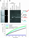
Fig. 1.
Fig. 1.
Fluorescent human Rad51 protein. (A Left) A Coomassie-stained gel showing wt Rad51, the cysteine minus Rad51 mutant, and A11C Rad51, which has a single surface cysteine. All three proteins were subjected to the same labeling and purification procedure (Supporting Materials and Methods, which is published as supporting information on the PNAS web site). (A Right) A fluorescence image of the same gel is shown. (B) Unlabeled wt or fluorescently tagged Rad51 was assembled onto circular single-stranded DNA and reacted with homologous linear duplex DNA. The resulting reaction products were deproteinized, resolved on an agarose gel, and stained with ethidium bromide. (C) ATPase assays with wt Rad51, unlabeled A11C Rad51, and the fluorescently tagged version of A11C Rad51.
Fig. 2.
Fig. 2.
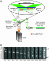
Human Rad51 slides on dsDNA. (A) Biotinylated λ-DNA molecules were tethered to a sample chamber surface protected with a lipid bilayer. Hydrodynamic force was used to extend the DNA molecules parallel to the surface. A 532-nm laser provided illumination, and data were collected by using an electron-multiplying charge-coupled device (CCD). (B) An image sequence showing Rad51 movement on an individual DNA molecule. Five nanomolar fluorescent Rad51 and 2 mM ATP were injected into the sample chamber, and images collected at 40-sec intervals for the duration of the experiment. The DNA is oriented vertically in the center of each frame. Off-axis fluorescent signals are due to protein molecules nonspecifically adhered to the surface and serve as stationary reference points. The numbers at the bottom show elapsed time, arrows indicate the direction of flow and highlight the movement of Rad51, and the tethered (T) and free (F) ends of the DNA molecule are indicated (refer to Movie 1 for a movie of Rad51 sliding).
Fig. 3.
Fig. 3.
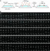
Human Rad51 stops sliding at the free ends of the DNA. (A) Shows a depiction of the tethered DNA molecules and their response to changes in hydrodynamic force. (B) Tethered DNA molecules were assembled into an aligned array by using a combination of hydrodynamic force and microscale barriers to lipid diffusion. The DNA molecules were stained with YOYO1 and illuminated at 488 nm. In the presence of flow, all of the molecules can be view across their full contour lengths. In the absence of flow, the DNA molecules experience an increase in conformational entropy and diffuse out of the evanescent field. The free (F) and tethered (T) ends of the DNA molecules are indicated. (C) A DNA array bound by fluorescent Rad51 is shown. Each fluorescent spot within the array corresponds to a single-protein complex sliding down a DNA molecule, and the accumulation of Rad51 at the free end of the DNA is evident as a fluorescent “line” of protein extending across the array. In the absence of flow, the DNA molecules and the DNA-bound proteins diffuse out of view, confirming that they did not nonspecifically adhere to the lipid bilayer. Each image represents a single 100-msec frame taken from a real-time video (Movie 2). (Scale bar: 10 μm.)
Fig. 4.
Fig. 4.
The movement of Rad51 on dsDNA occurs via a 1D-diffusion mechanism. (A) λ-DNA was tethered by both ends to a fused silica surface coated with a supported lipid bilayer. Rad51 and ATP were then injected into the sample chamber and allowed a brief period for binding. Unbound protein and ATP were then flushed from the sample chamber. (B) An image sequence showing the movement of Rad51 on dsDNA in the absence of flow force and ATP. Images were collected at 20-sec intervals for a total of 33 min, and the individual Rad51 complexes on the DNA are highlighted with arrowheads. Three different complexes are highlighted, although two occasionally come into close proximity with one another and can no longer be spatially resolved. Fluorescent signals not aligned with the axis of the DNA are due to proteins adsorbed to the sample chamber surface and serve as stationary reference points (see also Movie 4).
Fig. 5.
Fig. 5.
Characteristics of diffusing complexes. (A) A graphical representation of the y displacement (i.e., parallel to the helical axis of the tethered DNA) is shown of three typical Rad51 complexes bound to dsDNA molecules monitored over a period of 124 sec. Measurements were made with double-tethered DNA in the absence of buffer flow, images were collected at 8.3 frames per sec, the positions of the protein complexes were measured by fitting the images to a 2D-Gaussian function, and movement was measured as the frame-to-frame change in position for each individual protein complex. (B) Shows the x displacement (i.e., perpendicular to the helical axis of the tethered DNA) for the three diffusing protein complexes. Time-dependent variations in the amplitude reflect local flexibility of the extended DNA molecules as well as changes in the signal-to-noise ratio as the fluorophores bleach over time. (C) The mean squared displacement for these three complexes was plotted as a function of time interval for a period up to 12 sec. ○, calculated data points; solid lines, linear fits to the data points. (D) A graph of the total distance versus time for the same three Rad51 complexes is shown. In D, the points on the graph represent the absolute value of the change in position; direction of movement is not implied. Each trace represents the total distance traversed by a single Rad51 complex during the indicated time interval. Like colors correspond to the same Rad51 particles in each of the four graphs.

References

    1. Sung, P., Krejci, L., Van Komen, S. & Sehorn, M. G. (2003) J. Biol. Chem. 278, 42729–42732. - PubMed
    1. Cromie, G. A., Connelly, J. C. & Leach, D. R. F. (2001) Mol. Cell 8, 1163–1174. - PubMed
    1. Symington, L. S. (2002) Microbiol. Mol. Biol. Rev. 66, 630–670. - PMC - PubMed
    1. Tsuzuki, T., Fujii, Y., Sakumi, K., Tominaga, Y., Nakao, K., Sekiguchi, M., Yoshimura, Y. & Morita, T. (1996) Proc. Natl. Acad. Sci. USA 93, 6236–6240. - PMC - PubMed
    1. Khanna, K. K. & Jackson, S. P. (2001) Nat. Genet. 27, 247–254. - PubMed

Publication types