. 2006 May 15;396(1):31-9.
doi: 10.1042/BJ20051916.

Localization of GRP78 to mitochondria under the unfolded protein response

Affiliations

Localization of GRP78 to mitochondria under the unfolded protein response

Fang-Chun Sun et al. Biochem J. .

Abstract

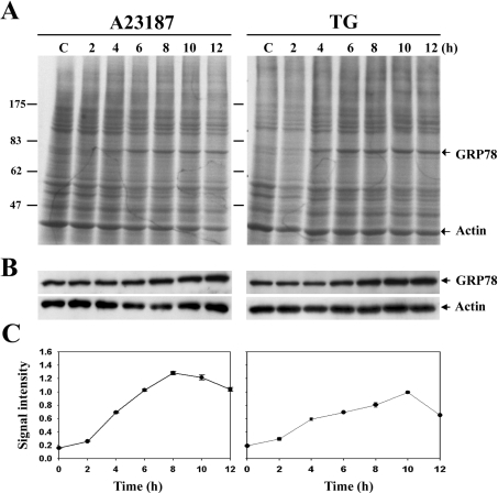
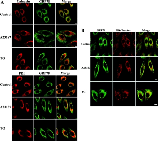
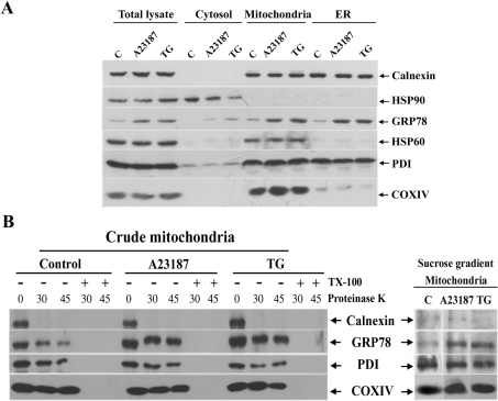
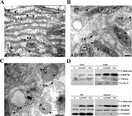
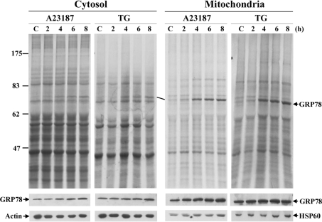
The ubiquitously expressed molecular chaperone GRP78 (78 kDa glucose-regulated protein) generally localizes to the ER (endoplasmic reticulum). GRP78 is specifically induced in cells under the UPR (unfolded protein response), which can be elicited by treatments with calcium ionophore A23187 and sarcoplasmic/endoplasmic reticulum Ca2+-ATPase inhibitor TG (thapsigargin). By using confocal microscopy, we have demonstrated that GRP78 was concentrated in the perinuclear region and co-localized with the ER marker proteins, calnexin and PDI (protein disulphide-isomerase), in cells under normal growth conditions. However, treatments with A23187 and TG led to diminish its ER targeting, resulting in redirection into a cytoplasmic vesicular pattern, and overlapping with the mitochondrial marker MitoTracker. Cellular fractionation and protease digestion of isolated mitochondria from ER-stressed cells suggested that a significant portion of GRP78 is localized to the mitochondria and is protease-resistant. Localizations of GRP78 in ER and mitochondria were confirmed by using immunoelectron microscopy. In ER-stressed cells, GRP78 mainly localized within the mitochondria and decorated the mitochondrial membrane compartment. Submitochondrial fractionation studies indicated further that the mitochondria-resided GRP78 is mainly located in the intermembrane space, inner membrane and matrix, but is not associated with the outer membrane. Furthermore, radioactive labelling followed by subcellular fractionation showed that a significant portion of the newly synthesized GRP78 is localized to the mitochondria in cells under UPR. Taken together, our results indicate that, at least under certain circumstances, the ER-resided chaperone GRP78 can be retargeted to mitochondria and thereby may be involved in correlating UPR signalling between these two organelles.


Figures

Figure 1
Figure 1. Induction of GRP78 in ER-stressed 9L RBT cells
(A) Cells were treated with A23187 and TG for the durations indicated and were metabolically labelled with [35S]methionine for 1 h before being harvested. Protein samples prepared from equivalent amounts of cells were resolved by SDS/PAGE and were visualized by autoradiography. Molecular-mass sizes are given in kDa. C, control. (B) Protein samples were separated by SDS/PAGE and were subjected to Western blotting with anti-GRP78 and anti-actin antibodies. (C) Relative signal intensities of GRP78 in the autoradiography were determined by densitometric scanning and were normalized for actin signals. Results are means±S.E.M. for three independent experiments.
Figure 2
Figure 2. Intracellular distribution of GRP78 before and after ER stress
(A) Cells were stained with anti-calnexin or anti-PDI antibodies followed by Cy5-conjugated secondary antibodies, and anti-GRP78 antibodies followed by FITC-conjugated secondary antibodies. The yellow signals in the merge images indicate the co-localization of GRP78 and ER marker protein. Scale bar, 10 μm. (B) Cells were incubated with MitoTracker, fixed and stained with anti-GRP78 antibodies followed by FITC-conjugated secondary antibodies. The yellow signals in the merge images indicate the co-localization of GRP78 and mitochondria. Scale bar, 10 μm.
Figure 3
Figure 3. Subcellular localization of GRP78 in normal and ER-stressed cells
Cells were fractionated by differential centrifugation, and the samples were analysed using Western blotting using antibodies against the indicated proteins. HSP90, HSP60 or COXIV, and calnexin or PDI were used as specific marker proteins for cytosol, mitochondria and ER respectively. C, control. (A) The amounts of protein loaded were 20 μg for total lysate, cytosol, crude mitochondria and ER fractions. (B) Proteinase K digestion of crude mitochondrial fractions (20 μg) for 30 or 45 min in the presence or absence of detergent (Triton X-100). Right-hand panel, purified mitochondria from sucrose-density gradient.
Figure 4
Figure 4. Submitochondrial localization of GRP78 in normal and ER-stressed cells
(A) Control, (B) A23187-treated and (C) TG-treated cells were processed for immunoelectron microscopy. Rabbit polyclonal anti-GRP78 antibodies and colloidal-gold-conjugated goat anti-rabbit IgG were used as the primary and secondary antibodies respectively. Arrows point to the localization of GRP78. Scale bars, 200 nm. M, mitochondrion. (D) The crude mitochondrial fractions were fractionated further into outer membrane (OM), IMS, inner membrane (IM) and matrix as described in the Materials and methods section. The protein samples were subjected to Western blotting. VDAC, cytochrome c (Cyto C), COXIV and HSP60 were used as markers for the submitochondrial fractions. Calnexin was used as a marker for the ER fraction.
Figure 5
Figure 5. Mitochondrial localization of newly synthesized GRP78
Cells were treated with A23187 or TG for different durations and then labelled with [35S]methionine for 1 h before being harvested. The cells were separated into cytosolic and mitochondrial fractions by differential centrifugation as described in the Materials and methods section. Each extract (20 μg of protein) was analysed by SDS/10% PAGE and was visualized by autoradiography. Western blotting with the antibodies against actin and HSP60 were used as markers for the cytosol and mitochondrial fractions respectively. Molecular-mass sizes are given in kDa. C, control.

References

    1. Brostrom M. A., Brostrom C. O. Calcium dynamics and endoplasmic reticular function in the regulation of protein synthesis: implications for cell growth and adaptability. Cell Calcium. 2003;34:345–363. - PubMed
    1. Sitia R., Braakman I. Quality control in the endoplasmic reticulum protein factory. Nature (London) 2003;426:891–894. - PubMed
    1. Meldolesi J., Pozzan T. The heterogeneity of ER Ca2+ stores has a key role in nonmuscle cell signaling and function. J. Cell Biol. 1998;142:1395–1398. - PMC - PubMed
    1. Berridge M. J. The endoplasmic reticulum: a multifunctional signaling organelle. Cell Calcium. 2002;32:235–249. - PubMed
    1. Kaufman R. J., Scheuner D., Schroder M., Shen X., Lee K., Liu C. Y., Arnold S. M. The unfolded protein response in nutrient sensing and differentiation. Nat. Rev. Mol. Cell Biol. 2002;3:411–421. - PubMed

Publication types