

Disaggregation and invasion of ovarian carcinoma ascites spheroids
- PMID: 16433903
- PMCID: PMC1397876
- DOI: 10.1186/1479-5876-4-6
Disaggregation and invasion of ovarian carcinoma ascites spheroids
Abstract
Background: Malignant ascites often develops in advanced stages of ovarian carcinoma, consisting of single and aggregated tumor cells, or spheroids. Spheroids have commonly been used as tumor models to study drug efficacy, and have shown resistance to some chemotherapies and radiation. However, little is known about the adhesive or invasive capabilities of spheroids, and whether this particular cellular component of the ascites can contribute to dissemination of ovarian cancer. Here, we examined the invasive ability of ascites spheroids recovered from seven ovarian carcinoma patients and one primary peritoneal carcinoma (PPC) patient.
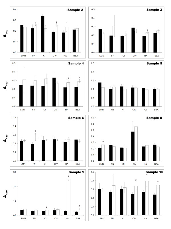
Methods: Ascites spheroids were isolated from patients, purified, and immunohistochemical analyses were performed by a pathologist to confirm diagnosis. In vitro assays were designed to quantify spheroid disaggregation on a variety of extracellular matrices and dissemination on and invasion into normal human mesothelial cell monolayers. Cell proliferation and viability were determined in each assay, and statistical significance demonstrated by the student's t-test.
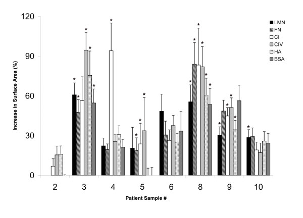
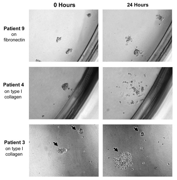
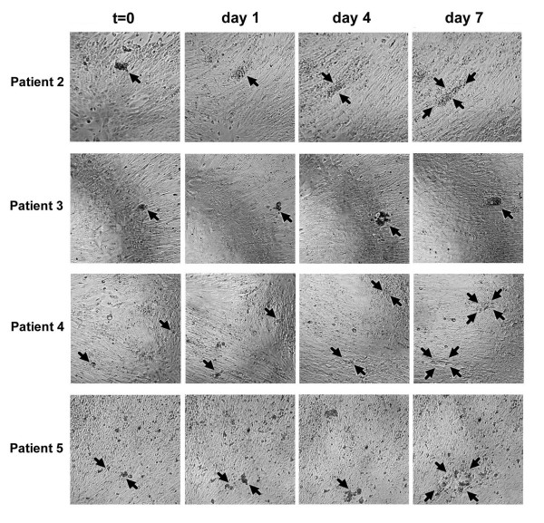
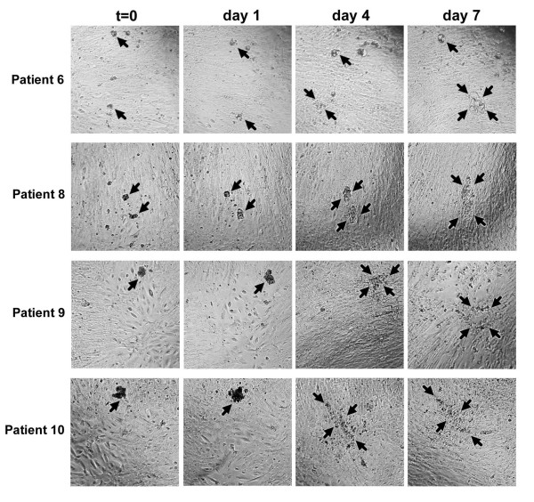
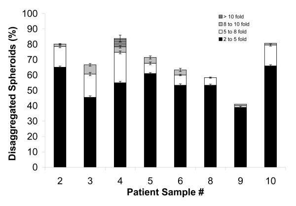
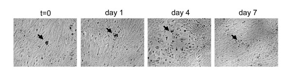
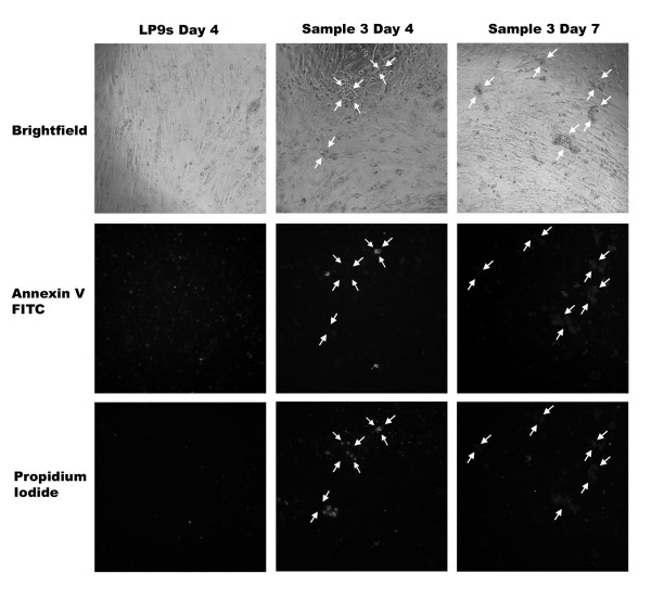
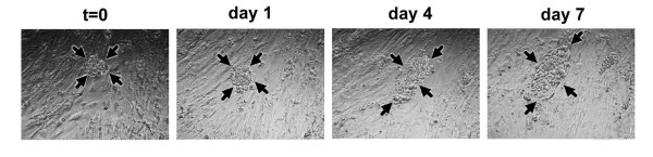
Results: Spheroids from all of the patients' ascites samples disaggregated on extracellular matrix components, with the PPC spheroids capable of complete disaggregation on type I collagen. Additionally, all of the ascites spheroid samples adhered to and disaggregated on live human mesothelial cell monolayers, typically without invading them. However, the PPC ascites spheroids and one ovarian carcinoma ascites spheroid sample occasionally formed invasive foci in the mesothelial cell monolayers, suggestive of a more invasive phenotype.
Conclusion: We present here in vitro assays using ascites spheroids that imitate the spread of ovarian cancer in vivo. Our results suggest that systematic studies of the ascites cellular content are necessary to understand the biology of ovarian carcinoma.
Figures
References
-
- Feldman GB, Knapp RC, Order SE, Hellman S. The role of lymphatic obstruction in the formation of ascites in a murine ovarian carcinoma. Cancer Research. 1972;32:1663–1666. - PubMed
-
- Feldman GB, Knapp RC. Lymphatic drainage of the peritoneal cavity and its significance in ovarian cancer. American Journal of Obstetrics and Gynecology. 1974;119:991–994. - PubMed
-
- Olson TA, Mohanraj D, Carson LF, Ramakrishnan S. Vascular permeability factor gene expression in normal and neoplastic human ovaries. Cancer Research. 1994;54:276–280. - PubMed
-
- Senger DR, Galli SJ, Dvorak AM, Perruzzi CA, Harvey VS, Dvorak HF. Tumor cells secrete a vascular permeability factor that promotes accumulation of ascites fluid. Science. 1983;21:983–985. - PubMed
Grants and funding
LinkOut - more resources
Full Text Sources
Other Literature Sources
Research Materials
