. 2006 Jan 31;103(5):1440-5.
doi: 10.1073/pnas.0510518103. Epub 2006 Jan 24.

Innate antiviral response targets HIV-1 release by the induction of ubiquitin-like protein ISG15

Affiliations

Innate antiviral response targets HIV-1 release by the induction of ubiquitin-like protein ISG15

Atsushi Okumura et al. Proc Natl Acad Sci U S A. .

Abstract

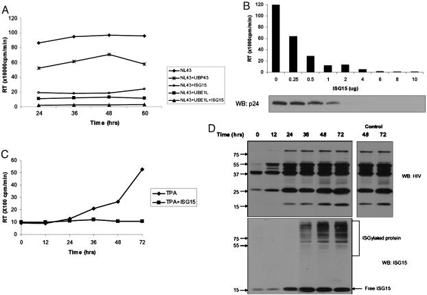
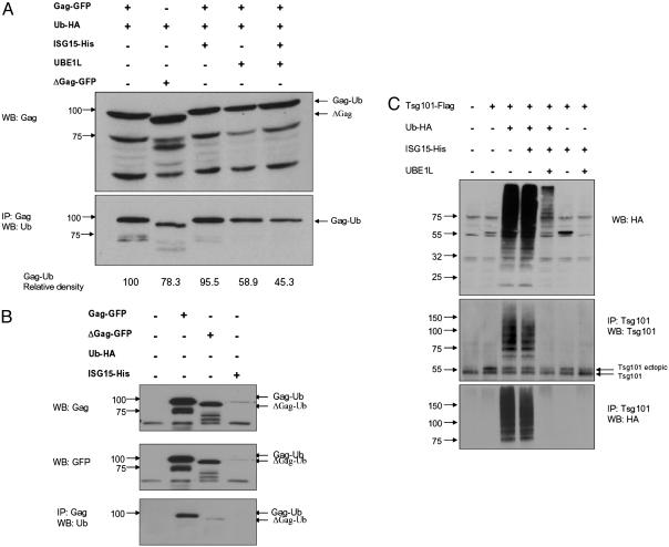
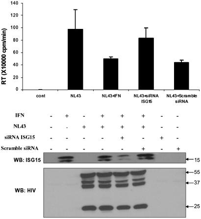
The goal of this study was to elucidate the molecular mechanism by which type I IFN inhibits assembly and release of HIV-1 virions. Our study revealed that the IFN-induced ubiquitin-like protein ISG15 mimics the IFN effect and inhibits release of HIV-1 virions without having any effect on the synthesis of HIV-1 proteins in the cells. ISG15 expression specifically inhibited ubiquitination of Gag and Tsg101 and disrupted the interaction of the Gag L domain with Tsg101, but conjugation of ISG15 to Gag or Tsg101 was not detected. The inhibition of Gag-Tsg101 interaction was also detected in HIV-1 infected, IFN-treated cells. Elimination of ISG15 expression by small interfering RNA reversed the IFN-mediated inhibition of HIV-1 replication and release of virions. These results indicated a critical role for ISG15 in the IFN-mediated inhibition of late stages of HIV-1 assembly and release and pointed to a mechanism by which the innate antiviral response targets the cellular endosomal trafficking pathway used by HIV-1 to exit the cell. Identification of ISG15 as the critical component in IFN-mediated inhibition of HIV-1 release advances the understanding of the IFN-mediated inhibition of HIV-1 replication and uncovers a target for the anti HIV-1 therapy.


Figures

Fig. 1.
Fig. 1.
ISG 15 inhibits HIV-1 replication. (A) HIV-1 (NL43) proviral DNA and plasmids expressing ISG15, UBE1L, or UBP43 (1 μg) were cotransfected to 293T cells. Supernatants were collected at indicated times after transfection, and the levels of released virus were analyzed by RT assay. (B) 293T cells were cotransfected with plasmids expressing NL43 (2 μg) and ISG15 (0 to 10 μg), and 48 h after transfection; supernatants were collected for RT assay. The purified virions (Supporting Text) were analyzed by immune blotting with p24 antibodies. The levels of transfected DNA were kept constant by the inclusion of plasmids DNA. (C) U1.1 cells were first treated with TPA for 24 h and then infected with 301 ISG15 vector or 301 control vector that were VSV pseudotyped. At different times after TPA treatment, supernatants were collected and the virus levels determined by the RT assay. (D) Cell lysates from TPA-treated U1.1 cells, infected with 301-ISG15 viral vector (described in C) or the empty 301 vector, were analyzed at different times after TPA treatment by Western blot with HIV and ISG15 antibodies. The levels of Gag proteins in TPA-treated U1.1 cells infected with an empty 301 vector at 48 and 72 h are shown.
Fig. 2.
Fig. 2.
ISG15 inhibits ubiquitination of Gag and Tsg101. (A) 293T cells were cotransfected with plasmids expressing optimized Gag or its p6 deletion mutant ΔGag. When indicated, cells were also transfected with Ub-HA-, ISG15-, and UBE1L-expressing plasmids. Cell lysates were analyzed at 48 h after transfection by Western blot with Gag polyclonal antibody (Upper), or immunoprecipitated with Gag and the precipitates analyzed by Western blot with anti-Ub monoclonal antibody (Lower). The integrated density value of bands is based on the comparison with Gag-Ub (100%). (B) 293T cells were transfected with GFP-Gag and GFP-ΔGag plasmids and the presence of Gag in lysates of transfected cells was determine by immunoblotting with Gag, GFP, or Ub antibodies. (C) Cells were cotransfected with Tsg101, Ub-HA, and, when indicated, ISG15 and UBE1L expression plasmids. Cell lysates were analyzed 24 h after transfection by Western blot with HA antibody (Top), or immunoprecipitated with Tsg101 antibody, and the precipitates were analyzed by immunoblotting with Tsg101 antibody (Middle) or HA antibodies (Bottom). All of the analyses were done in two independent experiments.
Fig. 3.
Fig. 3.
ISG15 inhibits Gag-Tsg101 interaction. (A) 293T cells were cotransfected with NL43 proviral DNA and Tsg101 expressing plasmid and, when indicated, also with ISG15 and UBE1L or UBP43 plasmids. Cell lysates (1 mg), prepared 48 h after transfection, were immunoprecipitated with Tsg101 or Gag antibody. The precipitates were analyzed by Western blot with Gag- or Tsg101-specific antibodies. (B) 293T cells were cotransfected with NL43 proviral DNA and, and when indicated, also with Tsg101-, UBE1L-, and UbcH8-expressing plasmids. Twenty-four hours after transfection, cells were treated with IFN-α (500 units/ml) for 48 h and then cell lysates (1 mg) were immunoprecipitated with Tsg101 antibody. The precipitates were analyzed by Western blot with Gag antibody (Top) or Tsg101 antibody (Middle). Presence of Gag in the cell lysates of transfected cells was determined by Western blot (Bottom). All of the analysis was done in two independent experiments.
Fig. 4.
Fig. 4.
Rescue of the IFN-mediated inhibition of HIV-1 release by ISG15 siRNA. 293T cells were cotransfected with NL43, ISG15 siRNA, or scrambled siRNA, and 48 h after transfection, cells were treated/untreated with IFN-α (500 units/ml) for 24 h. HIV-1 virions in the supernatants were analyzed by the RT assay. Cell lysates (20 μg of proteins) were analyzed by Western blot with ISG15 monoclonal antibody and the presence of Gag proteins was detected by immunoblotting with human HIV-1 antiserum (Tulpin). As a control, cells were transfected either with 10 nmol of siRNA or scrambled siRNA, and 48 h after transfection, cell lysates were analyzed by Western blot with ISG15 monoclonal antibodies.

References

    1. de Veer, M. J., Holko, M., Frevel, M., Walker, E., Der, S., Paranjape, J. M., Silverman, R. H. & Williams, B. R. (2001) J. Leukoc. Biol. 69, 912-920. - PubMed
    1. Stark, G. R., Kerr, I. M., Williams, B. R., Silverman, R. H. & Schreiber, R. D. (1998) Annu. Rev. Biochem. 67, 227-264. - PubMed
    1. Nyman, T. A., Matikainen, S., Sareneva, T., Julkunen, I. & Kalkkinen, N. (2000) Eur. J. Biochem. 267, 4011-4019. - PubMed
    1. Farrell, P. J., Broeze, R. J. & Lengyel, P. (1979) Nature 279, 523-525. - PubMed
    1. Zhao, C., Beaudenon, S. L., Kelley, M. L., Waddell, M. B., Yuan, W., Schulman, B. A., Huibregtse, J. M. & Krug, R. M. (2004) Proc. Natl. Acad. Sci. USA 101, 7578-7582. - PMC - PubMed

Publication types

MeSH terms

LinkOut - more resources