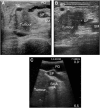
Refining the role of laparoscopy and laparoscopic ultrasound in the staging of presumed pancreatic head and ampullary tumours
- PMID: 16434983
- PMCID: PMC2361120
- DOI: 10.1038/sj.bjc.6602919
Refining the role of laparoscopy and laparoscopic ultrasound in the staging of presumed pancreatic head and ampullary tumours
Abstract
Laparoscopy and laparoscopic ultrasound have been validated previously as staging tools for pancreatic cancer. The aim of this study was to identify if assessment of vascular involvement with abdominal computed tomography (CT) would allow refinement of the selection criteria for laparoscopy and laparoscopic ultrasound (LUS). The details of patients staged with LUS and abdominal CT were obtained from the unit's pancreatic cancer database. A CT grade (O, A-F) of vascular involvement was recorded by a single radiologist. Of 152 patients, who underwent a LUS, 56 (37%) had unresectable disease. Three of 26 (12%) patients with CT grade O, 27 of 88 (31%) patients with CT grade A to D, 17 of 29 (59%) patients with CT grade E and all nine patients with CT grade F were found to have unresectable disease. In all, 24% of patients with tumours <3 cm were found to have unresectable disease. In those patients with tumours considered unresectable, local vascular involvement was found in 56% of patients and vascular involvement with metastatic disease in 17%, while 20% of patients had liver metastases alone and 5% had isolated peritoneal metastases. The remaining patient was deemed unfit for resection. Selective use of laparoscopic ultrasound is indicated in the staging of periampullary tumours with CT grades A to D.
Figures


References
-
- Ahmed NA, Kochman ML, Lewis JD, Kadish S, Morris JB, Rosato EF, Ginsberg GG (2001) Endosonography is superior to angiography in the preoperative assessment of vascular involvement among patients with pancreatic carcinoma. J Clin Gastroenterol 32: 54–58 - PubMed
-
- Engelken FJF, Bettschart V, Rahman MQ, Parks RW, Garden OJ (2003) Prognostic factors in the palliation of pancreatic cancer. Eur J Surg Oncol 29: 368–373 - PubMed
-
- Falconer JS, Fearon KC, Ross JA, Elton R, Wigmore SJ, Garden OJ, Carter DC (1995) Acute-phase protein response and survival duration of patients with pancreatic cancer. Cancer 75: 2077–2082 - PubMed
-
- Ishiguchi T, Ota T, Naganawa S, Fukatsu H, Itoh S, Ishigaki T (2001) CT and MR imaging of pancreatic cancer. Hepatogastroenterology 48: 923–927 - PubMed
MeSH terms
LinkOut - more resources
Full Text Sources
Medical

