

Early and late immunohistochemical and ultrastructural changes associated with functional impairment of the lachrymal gland following external beam radiation
- PMID: 16436114
- PMCID: PMC2517348
- DOI: 10.1111/j.0959-9673.2006.00456.x
Early and late immunohistochemical and ultrastructural changes associated with functional impairment of the lachrymal gland following external beam radiation
Abstract
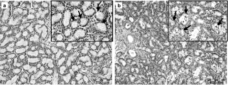
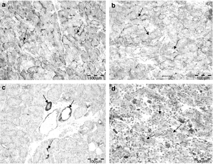
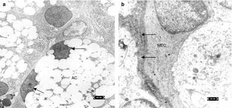
The aim of this study was to investigate scintigraphic, immunohistological and ultrastructural changes associated with radiation-induced dysfunction of the lachrymal gland in an established experimental animal model. Ten rabbits were randomized into two groups and used for the study; in the control as well as experimental group, the Schirmer-test, lachrymal gland scintigraphy, and immunohistological and ultrastructural investigations were carried out prior to irradiation and 72 h as well as 1 month after single-dose irradiation with 15 Gy. Seventy-two hours after irradiation, secretion reduction evaluated by the Schirmer-test was evident. At this phase, we could observe a decrease in the expression of alpha-SMA and a re-distribution of tenascin-C matrix. Ultrastructural changes of acinar and myoepithelial cells were noticed; simultaneously, disturbance in the primary (99m)TcO(4)- uptake as well as significant reduction of the lachrymal ejection fraction was assessed scintigraphically. These changes were still evident 1 month following irradiation but became less intensive. Single-dose irradiation with 15 Gy implicates a functional impairment of the lachrymal gland, which is associated with early immunohistological and ultrastructural alterations. These changes may represent objective surrogate parameters for radiogenic dysfunction and prerequisites for further investigations on radioprotection of lachrymal glands during radiotherapy of the periorbital region.
Figures
Similar articles
-
Pharmacological prevention of radiation-induced dry eye-an experimental study in a rabbit model.Graefes Arch Clin Exp Ophthalmol. 2007 Sep;245(9):1347-55. doi: 10.1007/s00417-007-0548-y. Epub 2007 Feb 21. Graefes Arch Clin Exp Ophthalmol. 2007. PMID: 17318564
-
[Establishment mice model of radiation-injured lacrimal gland and pathophysiological exploration].Zhonghua Yan Ke Za Zhi. 2014 Oct;50(10):758-65. Zhonghua Yan Ke Za Zhi. 2014. PMID: 25547579 Chinese.
-
Early immunohistochemical and functional markers indicating radiation damage of the parotid gland.Clin Oral Investig. 2004 Mar;8(1):30-5. doi: 10.1007/s00784-003-0249-9. Epub 2004 Jan 21. Clin Oral Investig. 2004. PMID: 14735331
-
The role of myoepithelial cells in the short-term radiogenic impairment of salivary glands. An immunohistochemical, ultrastructural and scintigraphic study.Anticancer Res. 2002 Nov-Dec;22(6C):4121-8. Anticancer Res. 2002. PMID: 12553042
-
Re-distribution of tenascin-C in the parotid acinar cells. An early marker of radiation-induced damage of salivary glands?Anticancer Res. 2004 Sep-Oct;24(5A):2841-6. Anticancer Res. 2004. PMID: 15517886
Cited by
-
First bite syndrome following surgical management of primary jugular foramen lesion: Clinical experience and review of the literature.Braz J Otorhinolaryngol. 2025 May 3;91(5):101629. doi: 10.1016/j.bjorl.2025.101629. Online ahead of print. Braz J Otorhinolaryngol. 2025. PMID: 40319535 Free PMC article.
-
First-bite syndrome in oncologic patients.Eur Arch Otorhinolaryngol. 2011 Aug;268(8):1241-1244. doi: 10.1007/s00405-011-1645-4. Epub 2011 May 26. Eur Arch Otorhinolaryngol. 2011. PMID: 21614466
-
Improvement of radiotherapy-induced lacrimal gland injury by induced pluripotent stem cell-derived conditioned medium via MDK and inhibition of the p38/JNK pathway.Int J Mol Sci. 2014 Oct 13;15(10):18407-21. doi: 10.3390/ijms151018407. Int J Mol Sci. 2014. PMID: 25314301 Free PMC article.
-
Application of Animal Models in Interpreting Dry Eye Disease.Front Med (Lausanne). 2022 Feb 1;9:830592. doi: 10.3389/fmed.2022.830592. eCollection 2022. Front Med (Lausanne). 2022. PMID: 35178415 Free PMC article. Review.
-
Review: The Lacrimal Gland and Its Role in Dry Eye.J Ophthalmol. 2016;2016:7542929. doi: 10.1155/2016/7542929. Epub 2016 Mar 2. J Ophthalmol. 2016. PMID: 27042343 Free PMC article. Review.
References
-
- Bartley GB, Gorman CA. Perspective – Part I: the Mayo Orbital Radiotherapy for Graves Ophthalmopathy (ORGO) study: lessons learned. Ophthal. Plast. Reconstr. Surg. 2002;18:170–172. - PubMed
-
- Eter N, Schuller H. External beam radiotherapy for age-related macular degeneration causes transient objective changes in tear-film function. Graefes Arch. Clin. Exp. Ophthalmol. 2001;239:923–926. - PubMed
-
- Garrett JR. The proper role of nerves in salivary secretion: a review. J. Dent. Res. 1987;66:387–397. - PubMed
-
- Gazda MJ, Schultheiss TE, Stephens LC, Ang KK, Peters LJ. The relationship between apoptosis and atrophy in the irradiated lachrimal gland. Int. J. Radiat. Oncol. Biol. Phys. 1992;24:693–697. - PubMed
-
- Hakim SG, Geerling G, Lauer I, Sieg P. Scintigraphic evaluation of lachrimal glands using a rabbit experimental model. Ophthalmic Res. 2002;34:254–257. - PubMed
MeSH terms
Substances
LinkOut - more resources
Full Text Sources
