. 2006 Apr 21;281(16):11260-70.
doi: 10.1074/jbc.M512885200. Epub 2006 Jan 25.

TRPM7 regulates cell adhesion by controlling the calcium-dependent protease calpain

Affiliations

TRPM7 regulates cell adhesion by controlling the calcium-dependent protease calpain

Li-Ting Su et al. J Biol Chem. .

Abstract

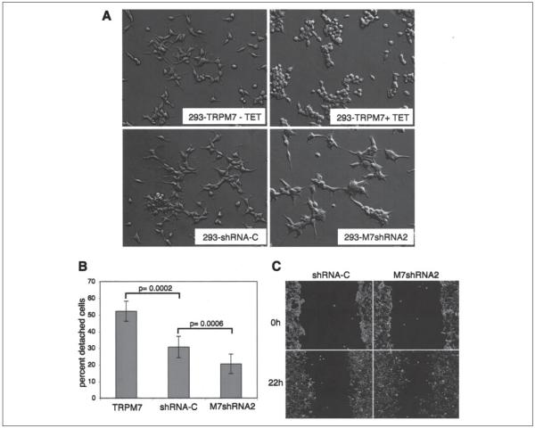
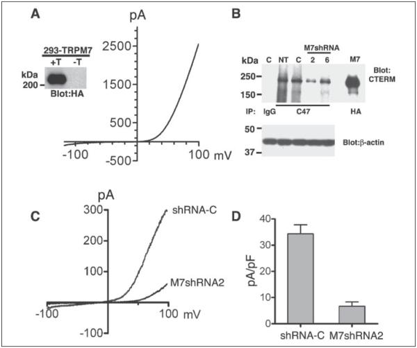
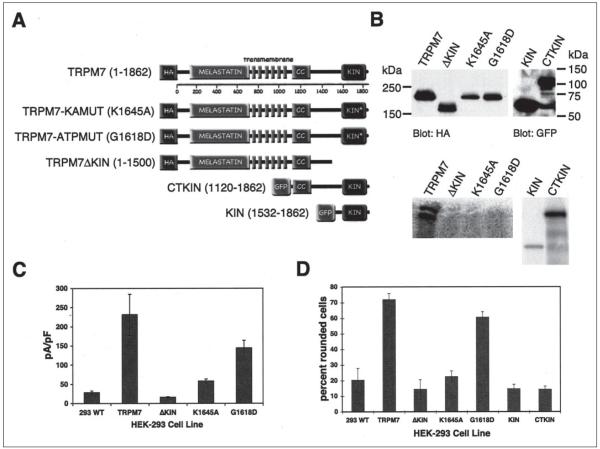
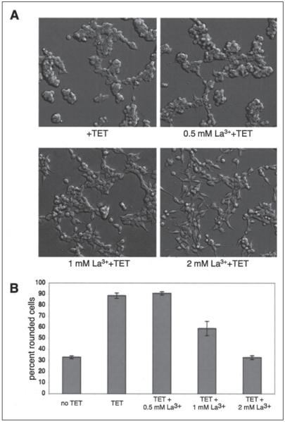
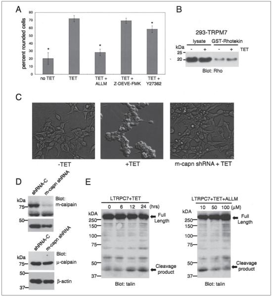
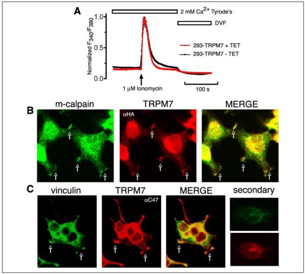
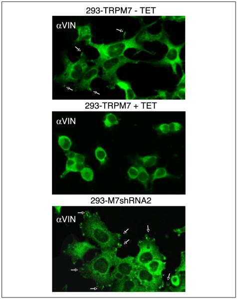
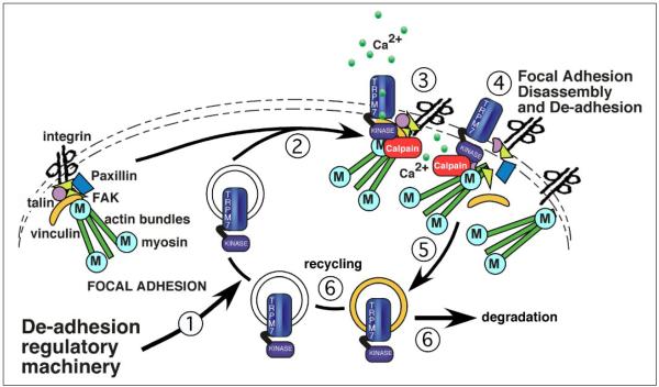
m-Calpain is a protease implicated in the control of cell adhesion through focal adhesion disassembly. The mechanism by which the enzyme is spatially and temporally controlled is not well understood, particularly because the dependence of calpain on calcium exceeds the submicromolar concentrations normally observed in cells. Here we show that the channel kinase TRPM7 localizes to peripheral adhesion complexes with m-calpain, where it regulates cell adhesion by controlling the activity of the protease. Our research revealed that overexpression of TRPM7 in cells caused cell rounding with a concomitant loss of cell adhesion that is dependent upon the channel of the protein but not its kinase activities. Knockdown of m-calpain blocked TRPM7-induced cell rounding and cell detachment. Silencing of TRPM7 by RNA interference, however, strengthened cell adhesion and increased the number of peripheral adhesion complexes in the cells. Together, our results suggest that the ion channel TRPM7 regulates cell adhesion through m-calpain by mediating the local influx of calcium into peripheral adhesion complexes.


Figures

FIGURE 1
FIGURE 1. TRPM7 regulates cell adhesion in HEK-293 cells
A, application of tetracycline (TET) induced expression of HA-TRPM7 and produced cell rounding of 293-TRPM7 cells. Knockdown of native TRPM7 in 293-M7shRNA2 cells produced cells that were more spread and had 50% longer extensions than a control cell line expressing a nonsilencing shRNA (293-shRNA-C). The same magnification was used in all four images. B, 293-M7shRNA2 cells adhered more strongly to the substratum than 293-shRNA-C or 293-TRPM7-expressing cells. Adhesion was measured using a trypsinization assay in which ~2.5 × 10⁶ cells were plated onto 60-mm Falcon tissue culture dishes for 24 h and then treated with 0.05% trypsin-EDTA for 4 min to stimulate cell detachment. The number of detached cells was counted using a hemocytometer and expressed as a percentage of the total number of cells on the plate. The error bars represent the standard error of the mean from seven independent experiments. p = 0.0002 and p = 0.0006 were compared with shRNA-C using the one-sided pair t test (α = 0.05). C, 293-M7shRNA2 cells migrated 56% more efficiently than 293-shRNA-C cells in a wound healing assay. The cells were outlined with white to enhance their visibility.
FIGURE 2
FIGURE 2. Characterization of 293-TRPM7 and 293-M7shRNA2 cells
A, expression of HA-tagged TRPM7 in the 293-TRPM7 cell line produced a whole cell current with a large outwardly rectifying conductance and small inward current at negative potentials. The inset shows tetracycline-induced (+T) expression of the 220-kDa channel by immunoprecipitation, SDS-PAGE, and Western blotting. −T, no tetracycline. B, cells stably expressing shRNAs targeting TRPM7 (shRNA-2 and -6; lanes 2 and 6, respectively) reduced TRPM7 expression compared with nontransfected (lane NT) cells or cells expressing a control shRNA (shRNA-C; lane C). Native TRPM7 was immunoprecipated with the anti-C47 antibody and detected with the anti-CTERM antibody. Rabbit IgG was used as an immunoprecipitation control. HA-tagged TRPM7 (lane M7) was immunoprecipitated with HA-agarose and detected with the anti-CTERM antibody as a migration control. Equivalent amounts of lysates were used for the immunoprecipitation study. A blot of β-actin is shown as a loading control. C, 293-M7shRNA2 cells express reduced amount of endogenous MagNUM/MIC current. D, the current-density of MagNUM/MIC current at +100 mV in 293-M7sRNA2 cells was reduced by 80% compared with 293-shRNA-C cells.
FIGURE 3
FIGURE 3. TRPM7 kinase activity is not required for changes in cell adhesion
A, one-dimensional structure of TRPM7 showing conserved melastatin domain followed by pore-forming transmembrane α-helices and a coiled-coil (CC) region that precedes the carboxyl-terminal kinase (KIN) domain. Substitution of K1645A or G1618D within the kinase domain rendered the kinase inactive. The TRPM7 variants shown were used to make individual 293 cell lines. B, top panel, Western blot showing expression levels of TRPM7 variants described in A. Bottom panel, autoradiograph showing immunokinase reactions demonstrate the capacity of the described TRPM7 variants to autophosphorylate. C, average current density at +100 mV from whole cell recordings of TRPM7 variant cell lines after 24 h of treatment with tetracycline. D, the capacity of 293 cells, 293-TRPM7, 293-TRPM7ΔKIN, 293-TRPM7-K1645A, 293-TRPM7-G1618D, 293-KIN, and 293-CTKIN to produce cell rounding upon induction of expression with tetracycline was scored manually (see “Experimental Procedures”). 293-TRPM7-G1618D cells, which express a kinase-inactive form of TRPM7 with high channel activity, produced cell rounding, whereas expression of the kinase domain alone (KIN) or the COOH terminus containing the kinase domain (CTKIN) did not. WT, wild type.
FIGURE 4
FIGURE 4. TRPM7 channel activity is required for cell rounding
A, application of La3+ to LTRPC7 cells treated with tetracycline (TET) inhibited cell rounding in a concentration-dependent manner. Full blockade was observed at 2 mm, a concentration that had been shown block TRPM7 inward current by 97% (1). B, quantization of the degree of cell rounding under the conditions depicted in A.
FIGURE 5
FIGURE 5. Calpain is required for TRPM7-mediated loss of cell adhesion
A, application of the calpain inhibitor ALLM (100 μm) to 293-TRPM7 cells treated with tetracycline (TET) for 24 h reduced TRPM7-induced cell rounding to a level similar to cells grown in the absence of tetracycline, as assessed by the scoring system described in the legend to Fig. 3. In contrast, treatment of cells with the caspase 3 inhibitor Z-DEVE-FMK (10 μm) or the Rho Kinase inhibitor (10 μm) had little effect. A χ2 test was employed to test differences in cell rounding between 293-TRPM7 cells treated with the different inhibitors. An asterisk indicates treatments that produced a decrease in cell rounding that was significantly different from 293-TRPM7 cells grown in tetracycline. B, a Rho activity assay shows that expression of TRPM7 (+TET) caused only a modest increase in Rho activity. C, expression of a shRNA targeting m-calpain blocked cell rounding and loss of adhesion in the LTRPC7 cell line. D, Western blot exhibiting the capacity of an shRNA targeting m-calpain to specifically reduce the protein levels of m-calpain in LTRPC7 cells. E, a Western blot using a monoclonal talin antibody that recognizes the head domain of talin was used to probe cell lysates from LTRPM7 cells treated with tetracycline for different durations. Expression of TRPM7 caused the cleavage of the focal adhesion protein talin into its head and rod domains. The head domain cleavage product migrated at 47 kDa. Application of increasing concentrations of the calpain inhibitor ALLM attenuated proteolysis of talin in response to TRPM7 expression.
FIGURE 6
FIGURE 6. Overexpression of TRPM7 does not cause calcium overload
A, normalized ratio of F340/F380 in 293-TRPM7 cells with or without tetracycline (TET) induction. The Ca2+ imaging traces were averaged from 24 cells for both groups. The experiment was repeated four times with similar results. No significant difference of the normalized ratio was observed when cells were constantly perfused with 2 mm Ca2+ Tyrode’s solution and divalent-free solution (DVF). The above results indicate that overexpression of TRPM7 does not cause calcium overload in 293-TRPM7 cells. B, heterologously expressed HA-TRPM7 colocalized with endogenous m-calpain to peripheral adhesion complexes (see arrows). Staining was performed on cells treated with tetracycline at concentrations below those required to cause full cell rounding. C, TRPM7 also colocalized with the focal adhesion protein vinculin to peripheral adhesion complexes. The nuclear fluorescence in the TRPM7 panels was from nonspecific secondary antibody staining, which was more easily detected because of the low expression level of TRPM7. The experiment was repeated four times with similar results.
FIGURE 7
FIGURE 7. TRPM7 regulates peripheral adhesion levels
293-TRPM7 grown in the absence of tetracycline extended protrusions with vinculin (αVIN) containing peripheral adhesions (see arrows) at their tips (top panel). Expression of TRPM7 (+TET) caused these cells to round and reduced the number of these cellular structures (middle panel). 293-M7shRNA2 cells, which had reduced levels of native TRPM7, were more spread and contained more peripheral adhesions, suggesting that TRPM7 controls cell adhesion by stimulating peripheral adhesion turnover. The experiment was repeated three times with similar results.
FIGURE 8
FIGURE 8. A working model of the role of TRPM7 in regulating cell adhesion
We hypothesize that recruitment of TRPM7 to areas of focal adhesion remodeling and turnover is under signal control (Step 1). The entry of TRPM7 into these cytoskeletal structures (Step 2) raises local calcium levels by virtue of increased calcium influx through the channel (Step 3). Calcium entry stimulates key molecules, such as m-calpain, to regulate focal adhesion disassembly. The role of the kinase domain is unknown, but it may be regulating key cytoskeletal events as well. Once focal adhesion turnover is completed, TRPM7 undergoes endocytosis (Step 5) and is then either recycled or degraded (Step 6).

References

    1. Runnels LW, Yue L, Clapham DE. Science. 2001;291:1043–1047. - PubMed
    1. Fleig A, Penner R. Novartis Found. Symp. 2004;258:248–266. - PubMed
    1. Nilius B, Voets T. Novartis Found Symp. 2004;258:140–159. 263–266. - PubMed
    1. Clapham DE. Nature. 2003;426:517–524. - PubMed
    1. Montell C, Birnbaumer L, Flockerzi V. Cell. 2002;108:595–598. - PubMed

Publication types

MeSH terms

LinkOut - more resources