. 2006 Apr;20(6):720-2.
doi: 10.1096/fj.05-5432fje. Epub 2006 Jan 25.

Premature aging-like phenotype in fibroblast growth factor 23 null mice is a vitamin D-mediated process

Affiliations

Premature aging-like phenotype in fibroblast growth factor 23 null mice is a vitamin D-mediated process

Mohammed S Razzaque et al. FASEB J. 2006 Apr.

Abstract

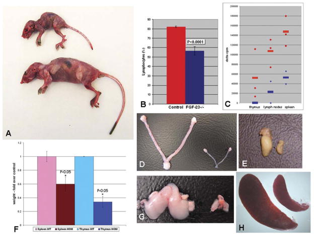
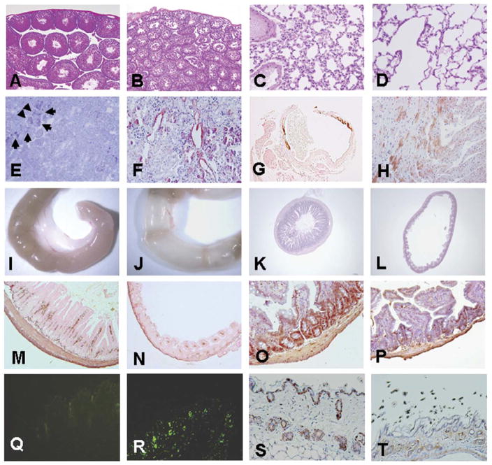
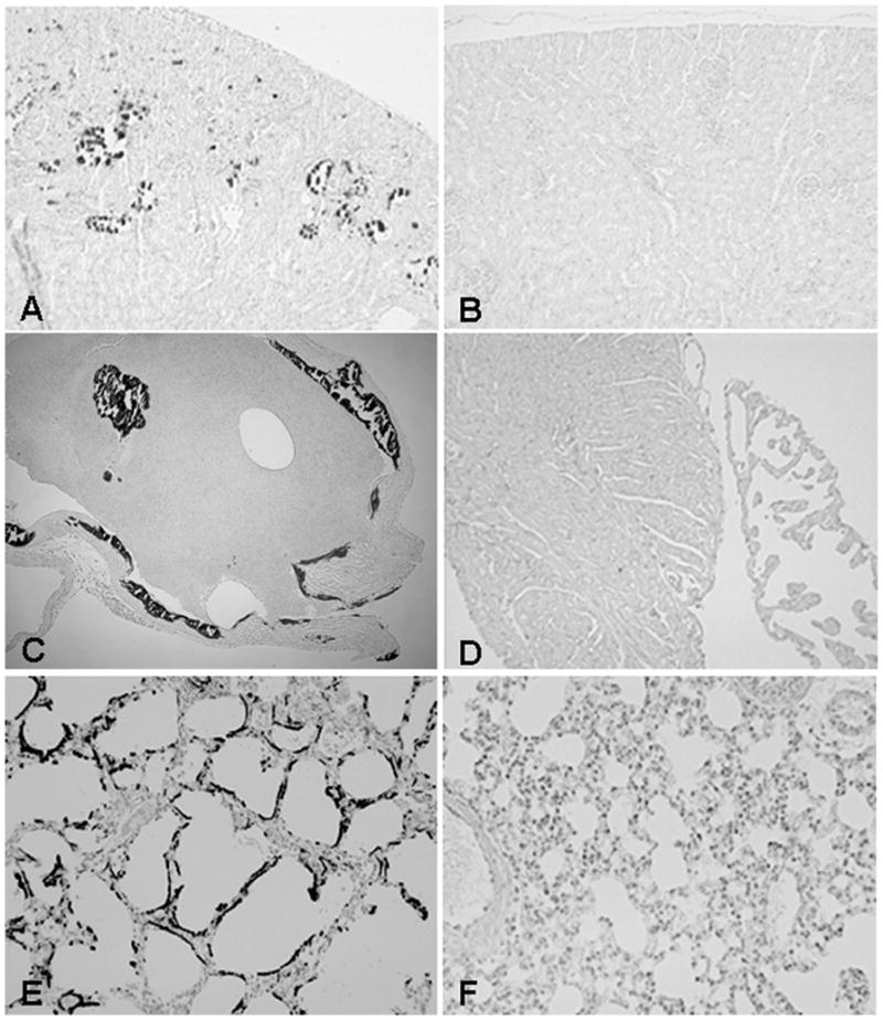
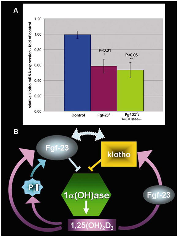
Fibroblast growth factor 23 null mice (Fgf-23-/-) have a short lifespan and show numerous biochemical and morphological features consistent with premature aging-like phenotypes, including kyphosis, severe muscle wasting, hypogonadism, osteopenia, emphysema, uncoordinated movement, T cell dysregulation, and atrophy of the intestinal villi, skin, thymus, and spleen. Furthermore, increased vitamin D activities in homozygous mutants are associated with severe atherosclerosis and widespread soft tissue calcifications; ablation of vitamin D activity from Fgf-23-/- mice, by genetically deleting the 1alpha(OH)ase gene, eliminates atherosclerosis and ectopic calcifications and significantly rescues premature aging-like features of Fgf-23-/- mice, resulting in prolonged survival of Fgf-23-/-/1alpha(OH)ase-/- double mutants. Our results indicate a novel role of Fgf-23 in developing premature aging-like features through regulating vitamin D homeostasis. Finally, our data support a new model of interactions among Fgf-23, vitamin D, and klotho, a gene described as being associated with premature aging process.


Figures

Figure 1
Figure 1
Premature aging features of Fgf-23−/− mice. A) Mutant mouse (top) shows small body size, reduced muscle mass, and kyphosis when compared with normal control littermate (bottom) at 6 wk of age. B) Number of lymphocytes in peripheral blood of Fgf-23−/− animals (n=9, blue bar) was significantly decreased (P<0.0001), when compared with controls (n=13, red bar). C) Mitogenic response of T cells isolated from thymus, lymph nodes, or spleen to concavanalin A. Shown are mean values (quadruplicates) of controls (n=3, red circles) and Fgf-23−/− animals (n=2, blue squares). Red and blue cross-lines represent mean of 12 measurements in controls (red lines) and 8 measurements in mutants (blue lines). D) Atrophy of mutant uterus and ovary (right) when compared with normal (left) at 6 wk of age leads to hypogonadism and infertility in females. E) Atrophied testes of Fgf-23−/− animals (right) also causes infertility in Fgf-23−/− males. F) Quantitative assessment of the weight of spleen and thymus (n=3, P<0.05). Data are presented as fold over control (=1) after each organ weight was normalized to total body weight of matching animal. Macroscopic picture of an atrophied thymus (G) and spleen (H) of Fgf-23−/− animals (right) when compared with normal control littermates (left).
Figure 2
Figure 2
Macroscopic and microscopic features of various organs at 6 wk of age (n=8). Hematoxilin- and eosin stained sections of wild-type (A) and Fgf-23−/− (B) testes and of control (C) and Fgf-23−/− (D) lungs (A–D: ×10). Mutant lung exhibits typical emphysematous features as observed during aging. E) Toluidine blue staining of an Fgf-23−/− kidney at 6 wk of age. Arrows depict kidney stones found only in the mutants. F) Congo-red staining indicates amyloid deposition found in small and medium sized arteries of the mutant kidney and heart (H). G) von Kossa staining demonstrates severe calcification of the aortic wall in Fgf-23−/− animals. Macroscopic image of small intestine of wild-type (I) and Fgf-23−/− animal (J) (n=6), showing ballooning of mutant intestine. Hematoxilin/eosin staining of cross sections of wild-type (K) and Fgf-23−/− littermate (L) (K and L: ×5). Reduced height of intestinal villi, and atrophy of intestinal mucosa are shown. Immunohistochemical evaluation by α-smooth muscle actin antibody staining presents dramatic reduction in vascularization and smooth muscle coat in intestine of mutant animals (N), when compared with normal littermates M). BrdU staining demonstrates striking diminution of proliferative cells in mutants (P) vs. wild-type (O) mice (M to P: ×10). R) Increased apoptosis in the mutant skin is visible by positive TUNEL staining mostly located in hair follicles when compared with normal (Q). BrdU labeling of the skin resulted in dramatic decrease in proliferative cells in Fgf-23−/− (T) vs. normal (S) littermate (Q to T: ×10).
Figure 3
Figure 3
Ectopic calcifications. Von Kossa staining of paraffin sections of kidney (A), heart (C; muscle and valves), and lung of Fgf-23−/− (E) animals demonstrating severe calcifications of these organs. Images presented in B, D, and F show complete elimination of abnormal calcification pattern in littermates that are deficient for both Fgf-23 and 1α(OH)ase gene (n=8; A to F: ×10).
Figure 4
Figure 4
A) Macroscopic image of a wild-type (WT), Fgf-23−/−, and Fgf-23−/−/1α(OH)ase−/− double mutant at ~6 wk of age. B) Body weight curve and survival curve (C) of control, Fgf-23−/− and Fgf-23−/−/1α (OH)ase−/− mice (n>15) showing gain of weight and prolonged lifespan in compound mutants. Hematoxilin/eosin staining of intestinal sections of wild-type (D), Fgf-23−/− (F), and Fgf-23−/−/1α(OH)ase−/− double knockout animals. Single Fgf-23−/− mice exhibit (E) severe atrophy of intestine, which is significantly improved by deletion of the 1α(OH)ase gene. Similar improvement was also noted in the skin section of Fgf-23−/−/1α(OH)ase−/− double knockout animals, compared with Fgf-23−/− mice; wild-type (G), Fgf-23−/− (H), and Fgf-23−/−/1α(OH)ase−/− (I) double knockout animals (D to I: ×10).
Figure 5
Figure 5
A) Quantitative real-time PCR was performed for klotho expression in kidney (n>3). Relative expression of klotho mRNA in kidneys of Fgf-23−/− animals (P<0.01) and in Fgf-23−/−/1α(OH)ase−/− double knockout animals (P<0.05) is significantly decreased when compared with controls. Data shown is the klotho mRNA expression as fold over control (=1). B) Schematic representation of a model showing the possible interactions among Fgf-23, vitamin D, and klotho. Fgf-23 activity is required for vitamin-D mediated expression of klotho.

References

    1. Johnson FB, Sinclair DA, Guarente L. Molecular biology of aging. Cell. 1999;96:291–302. - PubMed
    1. Dolle ME, Giese H, Hopkins CL, Martus HJ, Hausdorff JM, Vijg J. Rapid accumulation of genome rearrangements in liver but not in brain of old mice. Nat Genet. 1997;17:431–434. - PubMed
    1. Kirkwood TB, Austad SN. Why do we age? Nature. 2000;408:233–238. - PubMed
    1. de Boer J, Andressoo JO, de Wit J, Huijmans J, Beems RB, van Steeg H, Weeda G, van der Horst GT, van Leeuwen W, Themmen AP, et al. Premature aging in mice deficient in DNA repair and transcription. Science. 2002;296:1276–1279. - PubMed
    1. Delmas D, Jannin B, Latruffe N. Resveratrol: preventing properties against vascular alterations and aging. Mol Nutr Food Res. 2005;49:377–395. - PubMed

Publication types

MeSH terms

LinkOut - more resources