. 2006 Apr;17(4):1959-70.
doi: 10.1091/mbc.e05-07-0668. Epub 2006 Jan 25.

The Saccharomyces cerevisiae spindle pole body (SPB) component Nbp1p is required for SPB membrane insertion and interacts with the integral membrane proteins Ndc1p and Mps2p

Affiliations

The Saccharomyces cerevisiae spindle pole body (SPB) component Nbp1p is required for SPB membrane insertion and interacts with the integral membrane proteins Ndc1p and Mps2p

Yasuhiro Araki et al. Mol Biol Cell. 2006 Apr.

Abstract

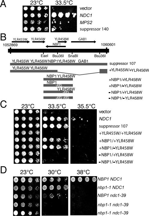
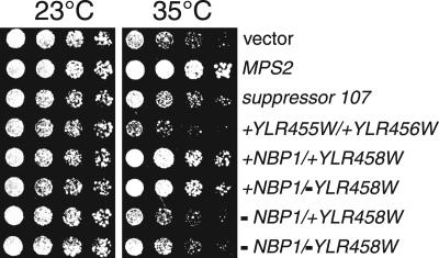
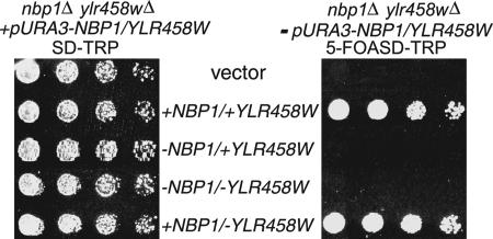
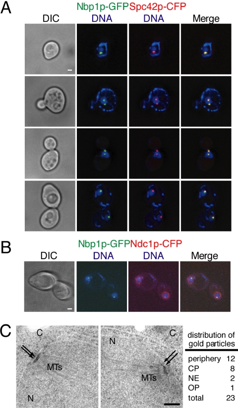
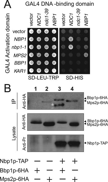
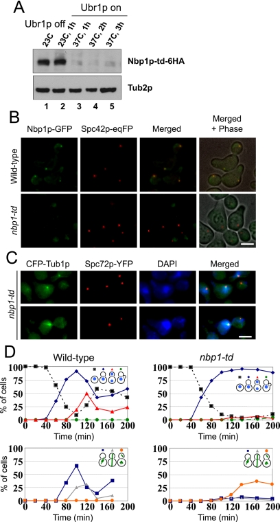
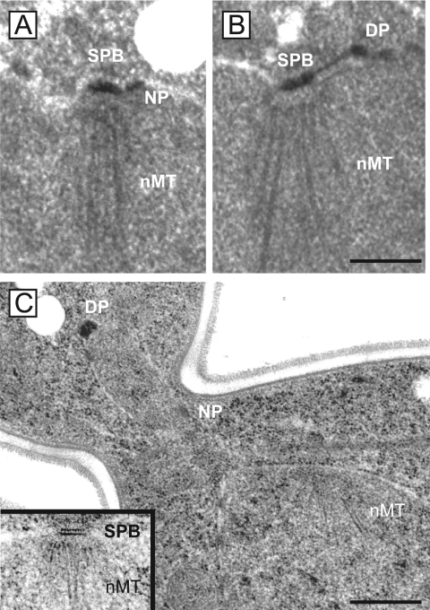
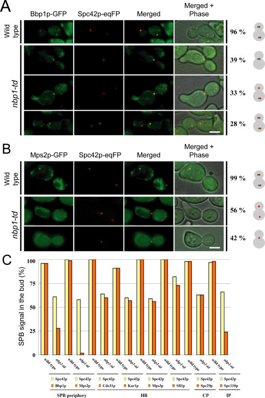
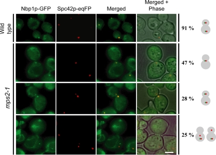
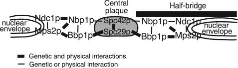
The spindle pole body (SPB) in Saccharomyces cerevisiae functions to nucleate and organize spindle microtubules, and it is embedded in the nuclear envelope throughout the yeast life cycle. However, the mechanism of membrane insertion of the SPB has not been elucidated. Ndc1p is an integral membrane protein that localizes to SPBs, and it is required for insertion of the SPB into the nuclear envelope during SPB duplication. To better understand the function of Ndc1p, we performed a dosage suppressor screen using the ndc1-39 temperature-sensitive allele. We identified an essential SPB component, Nbp1p. NBP1 shows genetic interactions with several SPB genes in addition to NDC1, and two-hybrid analysis revealed that Nbp1p binds to Ndc1p. Furthermore, Nbp1p is in the Mps2p-Bbp1p complex in the SPB. Immunoelectron microscopy confirmed that Nbp1p localizes to the SPB, suggesting a function at this location. Consistent with this hypothesis, nbp1-td (a degron allele) cells fail in SPB duplication upon depletion of Nbp1p. Importantly, these cells exhibit a "dead" SPB phenotype, similar to cells mutant in MPS2, NDC1, or BBP1. These results demonstrate that Nbp1p is a SPB component that acts in SPB duplication at the point of SPB insertion into the nuclear envelope.


Figures

Figure 1.
Figure 1.
MPS2 and NBP1 are dosage suppressors of ndc1-39. (A) Two-micron plasmids containing vector, NDC1 (Chial et al., 1999), MPS2 (McBratney and Winey, 2002), and suppressor 140 (contains DNA from chromosome IV, coordinates 366254-373702, which includes MPS2) were transformed into ndc1-39 cells (3446, Table 1). Cells were grown in YPD media to saturation at 23°C, then diluted to a density of 3 OD600 U/ml, and spotted onto YPD plates in fivefold serial dilutions. Plates were incubated for 4 d at 23 or 33.5°C. (B) A single clone containing DNA from coordinates 1052869-1060601 on chromosome XII was able to partially rescue the temperature sensitivity of ndc1-39 cells. This clone, suppressor 107, contained five ORFs. A clone containing only YLR455W and YLR456W (+YLR455W/+YLR456W) was created by cutting suppressor 107 with Bsu36I and religating the plasmid. A clone containing NBP1 and YLR458W (+NBP1/+YLR458W) was obtained by ligating a PCR-generated EaeI-SnaBI fragment into pRS202 (see Materials and Methods). The start codon of YLR458W was mutated so that only NBP1 is expressed in the +NBP1/-YLR458W plasmid (see Materials and Methods). The start codon of NBP1 was mutated so that only YLR458W is expressed in the -NBP1/+YLR458W plasmid. Both start codons of NBP1 and YLR458W were mutated to create the -NBP1/-YLR458W plasmid. (C) Two-micron URA3-based plasmids containing the indicated ORFs were transformed into ndc1-39 cells (3446, Table 1). Cells were grown and spotted onto YPD plates as in A. Plates were incubated for 4 d at 23, 33.5, or 35.5°C. (D) nbp1-1 cells are synthetically sick with ndc1-39 cells. Heterozygous diploids were generated by crossing nbp1-1 strain (YYS113-71, Table 1) to ndc1-39 strain (2711, Table 1) and then sporulated. Twenty tetrads were dissected and analyzed by spotting fivefold serial dilutions of cells at ∼5 OD600 U/ml on YPD plates. Plates were incubated at 23, 30, and 38°C for 2 d. The nbp1-1 ndc1-39 double mutant (3606, Table 1) was viable at 23°C but was unable to grow at 30°C, a temperature permissive for growth for the single mutant parents. Two isolates of nbp1-1 ndc1-39 double mutant are shown.
Figure 2.
Figure 2.
NBP1 also suppresses the growth defect of mps2-1. Two-micron plasmids containing the indicated ORFs (from Figure 1, A and C) were transformed into mps2-1 strain (1210, Table 1). Cells were grown and spotted onto YPD plates as in Figure 1A. Plates were incubated for 4 d at 23 or 35°C. NBP1 was able to rescue the growth defect of mps2-1 at 35°C, the semirestrictive temperature for mps2-1, but not at 37°C.
Figure 3.
Figure 3.
NBP1 is an essential gene. An nbp1Δ/ylr458wΔ strain (3445, Table 1) carrying a genomic deletion of NBP1 and YLR458W originally contained NBP1 and YLR458W on a CEN-URA3 plasmid. It was then transformed with the indicated ORFs (from Figure 1B) on CEN-TRP1 plasmids. The ability of the each transformed construct to rescue the lethality of the nbp1Δ/ylr458wΔ strain was tested by plating fivefold serial dilutions of cells on SD-TRP plates supplemented with 5-fluoroorotic acid (5-FOA; right panel) to select for the TRP1 plasmid and select against the URA3 plasmid. As a growth control, cells were spotted onto SD-TRP plates (left panel). Plates were incubated at 23°C for 4 d.
Figure 4.
Figure 4.
Nbp1p localizes to the periphery and the central plaque of SPBs. (A and B) Cells containing Nbp1p-GFP and Spc42p-CFP fusion proteins (3326, Table 1) or Nbp1p-GFP and Ndc1p-CFP (3322, Table 1) were grown in YPD media at 23°C, and processed for fluorescence microscopy the following day. DIC images are shown on the left column. Nbp1p-GFP (green) and Spc42p-CFP or Ndc1p-CFP (red) signals were detected by autofluorescence, and DNA was stained with DAPI (blue). Yellow color corresponds to colocalization signals. Examples of cells in various stages of the cell cycle are shown in A. Note that in B, Ndc1p-CFP has additional perinuclear signal corresponding to its known localization at the NPCs. Each fluorescent image shown was projected from 10 consecutive images taken at 0.1-μm intervals along the z-axis that have been deconvolved. Bar, 1 μm. (C) Localization of Nbp1p-GFP was examined by immunoelectron microscopy using anti-GFP antibodies and colloidal gold-conjugated secondary antibodies. Nbp1p-GFP cells (3310, Table 1) were grown to log-phase in YPD at 23°C and processed for immunoelectron microscopy the following day. The SPB is embedded in the nuclear envelope, which separates the nucleus (N) and cytoplasm (C). Microtubules (MTs) can be seen on the nuclear side of the SPB. Arrows indicate the positions of the gold particles that recognize Nbp1p-GFP. The two SPBs shown are from the same cell. A total of 11 SPBs were analyzed, and the distribution of gold particles to the central plaque region (CP), the outer plaque region (OP), the periphery of the SPB, and the nuclear envelope (NE) is indicated. Bar, 0.2 μm.
Figure 5.
Figure 5.
Nbp1p binds to both Ndc1p and the Mps2p-Bbp1p complex. (A) Two-hybrid analysis was done with PJ69-4a cells (Table 1) containing the indicated GAL4 activation domain fusions (rows) were crossed to PJ69-4α cells (Table 1) containing the indicated GAL4 DNA-binding domain fusions (columns), and diploids were selected on SD-LEU-TRP plates at 30°C (left). The ability of fusion proteins to interact was assayed by plating cells, which contain a version of HIS3 driven by the GAL1 promoter, on SD-HIS plates at 30°C (right). 30°C is the semipermissive temperature for strains containing ndc1-39 allele. Similar results were obtained at 23 and 37°C. (B) Coimmunoprecipitation of Bbp1p and Mps2p with Nbp1p. Lysates of BBP1-6HA (lane 1), MPS2-6HA (lane 2), NBP1-TAP BBP1-6HA (lane 3), or NBP1-TAP MPS2-6HA cells (lane 4) were subjected to TAP purification using IgG Sepharose (strains listed in Table 1, YAY167-170). IgG bound complexes were eluted with Tev protease. The eluates were analyzed by immunoblotting with anti-HA (12CA5) and anti-PrA antibodies. Lane 3 contains a degradation product of Bbp1p-6HA (asterisks). Mps2p-6HA (lane 2) bound weakly to IgG beads. However, the Mps2p-6HA signal in lane 4 was repeatedly stronger, indicating coimmunoprecipitation of Mps2p-6HA and Nbp1p-TAP.
Figure 6.
Figure 6.
nbp1-td cells are defective in mitotic spindle formation and chromosome segregation. (A) nbp1-td-6HA cells (YAY144, Table 1) were first grown at 23°C without expression of UBR1 (lane 1). Induction of UBR1 at 23°C did not affect Nbp1p-td-6HA protein levels as determined by immunoblotting with anti-HA (12CA5) antibodies (lane 2). However, upon shifting the cells to 37°C Nbp1p-td-6HA was rapidly degraded after 1, 2, or 3 h in the presence of UBR1 (lanes 3-5, respectively). An immunoblot using anti-Tub2p antibodies is shown as a protein loading control. (B) Nbp1p-td-GFP is not associated with SPBs. NBP1-GFP SPC42-eqFP (YAY157, Table 1) and nbp1-td-GFP SPC42-eqFP (YAY158, Table 1) cells were synchronized at 23°C using α-factor. Cells were then shifted to 37°C with UBR1 induction. We confirmed that Nbp1p-td-GFP was degraded upon shifting cells to 37°C by immunoblotting with anti-GFP antibodies. Consistently, no Nbp1p-td-GFP fluorescence signal was observed at SPBs. Bar, 5 μm. (C) nbp1-td cells fail to assemble a mitotic spindle. Wild-type and nbp1-td cells with CFP-TUB1 SPC72-YFP (SHM1097 and SHM1098, Table 1) were synchronized with α-factor in G1-phase of the cell cycle. α-factor was removed by washing the cells (t = 0). Samples were taken every 20 min. Fixed wild-type and nbp1-td cells were stained with DAPI and analyzed 140 min after release by fluorescence microscopy. (D) Quantification of C. More than 100 cells were counted per time point. nbp1-td cells arrested with a large bud with only one DAPI-staining region located in the mother cell body close to the bud neck (top panel, blue line). CFP-tubulin fluorescence revealed that >95% of the large budded nbp1-td cells failed to form a proper mitotic spindle. About 40% of nbp1-td cells had a SPB signal in the bud (bottom panel, orange line). This SPB organized 1-2 cytoplasmic but no nuclear microtubules. In these panels, percentages of total cells (budded or not) are reported.
Figure 7.
Figure 7.
nbp1-td cells fail to insert the new SPB into the nuclear envelope. Synchronized wild-type and nbp1-td cells (GPY658 and SHM1043, Table 1) were prepared for thin-section electron microscopy 1 h after shifting cells to 37°C. Serial sections of 20 cells were analyzed which spanned the entire nucleus. (A) All wild-type cells had a duplicated and separated SPB (only one of the two SPB is shown). (B) In contrast, 19/20 nbp1-td cells showed the mother SPB (SPB) associated through the bridge structure with the duplication plaque (DP). Nuclear microtubules (nMT) were only associated with the mother SPB but not the duplication plaque. One nbp1-td cell assembled a bipolar spindle. In this cell, nbp1p-td was probably not completely degraded. (C) A nbp1-td cell with the “dead pole” in the bud on an extension of the nuclear envelope. The nuclear microtubules mark the position of the mother SPB, which was found in a different section and shown in the inset at the same magnification as the cell. The cells in A and B were prepared by glutaraldehyde fixation, and the cell in C was prepared by high-pressure freezing and freeze substitution (see Materials and Methods). DP, duplication plaque (or “dead pole” in C); nMT, nuclear microtubules; NP, nuclear pore. Bars, 0.3 μm.
Figure 8.
Figure 8.
Bbp1p and Mps2p are not associated with the new SPB of nbp1-td cells. (A and B) Wild-type and nbp1-td cells with SPC42-eqFP and GFP-tagged SPB components (Table 1) were synchronized with α-factor and analyzed by fluorescence microscopy 135 min after release. Images of large-budded wild-type and nbp1-td cells expressing either BBP1-GFP (A; YAY075 and YAY074, Table 1) or MPS2-GFP (B; YAY093 and YAY101, Table 1) are shown. (A) Most large budded NBP1 (96%) and nbp1-td cells (61%) showed one SPB signal in the mother cell and one in the bud (n > 100). The remainder of large-budded nbp1-td cells showed only one Spc42p-eqFP SPB signal in the mother cell. Only about half of the large-budded nbp1-td cells that had two SPB signals contained Bbp1p in the new SPB in the bud. (B) In all nbp1-td cells, Mps2p failed to bind to the motherand daughter SPBs. Bar, 5 μm. (C) Wild-type and nbp1-td cells were synchronized as in A and B. Large-budded cells (n > 100) were scored for two SPB signals. On average, ∼60% of large budded nbp1-td cells showed Spc42p-eqFP signal in the mother cell and in the bud. Of these cells, for example, only 28% of cells carried a Bbp1p-GFP signal in the bud. An even stronger reduction of the SPB signal in the bud was observed for Spc110p and Mps2p, whereas Cdc31p, Kar1p, Mps3p, Sfi1p, and Spc29p behaved similar to Spc42p. HB, half-bridge; CP, central plaque; IP, inner plaque.
Figure 9.
Figure 9.
Localization of Nbp1p with SPBs is dependent on Mps2p. MPS2 (wild-type) and mps2-1 cells with NBP1-GFP SPC42-eqFP (YAY133 and YAY120, Table 1) were synchronized with α-factor at 23°C. After washing (t = 0), cells were shifted to 37°C and analyzed after 135 min by fluorescence microscopy for localization of Nbp1p-GFP at SPBs (marked by Spc42p-eqFP). Bar, 5 μm.
Figure 10.
Figure 10.
Model of Nbp1p interactions at the SPB. The relative positions of Nbp1p, Ndc1p, Mps2p, and Bbp1p between the nuclear envelope and the SPB's central plaque. The cytoplasm and the nucleoplasm are oriented on the top and the bottom of the nuclear envelope, respectively. For simplicity, only the central plaque components Spc42p and Spc29p and the half-bridge of the SPB are shown. Ndc1p and Mps2p are integral membrane proteins at the nuclear periphery of the SPB where they interact with Nbp1p. Nbp1p also binds to Mps2p-Bbp1p complex.

References

    1. Adams, I. R., and Kilmartin, J. V. (1999). Localization of core spindle pole body (SPB) components during SPB duplication in Saccharomyces cerevisiae. J. Cell Biol. 145, 809-823. - PMC - PubMed
    1. Adams, I. R., and Kilmartin, J. V. (2000). Spindle pole body duplication: a model for centrosome duplication? Trends Cell Biol. 10, 329-335. - PubMed
    1. Ausubel, F. M., Brent, R., Kingston, R. E., Moore, D. D., Seidman, J. G., Smith, J. A., and Struhl, K. (1998). Current Protocols in Molecular Biology, New York: John Wiley & Sons.
    1. Boeke, J. D., Trueheart, J., Natsoulis, G., and Fink, G. R. (1987). 5-Fluoroorotic acid as a selective agent in yeast molecular genetics. Methods Enzymol. 154, 164-175. - PubMed
    1. Byers, B., and Goetsch, L. (1975). Behavior of spindles and spindle plaques in the cell cycle and conjugation of Saccharomyces cerevisiae. J. Bacteriol. 124, 511-523. - PMC - PubMed

Publication types

MeSH terms