. 2006 Jan 25;26(4):1107-16.
doi: 10.1523/JNEUROSCI.3970-05.2006.

Loss of p53 induces changes in the behavior of subventricular zone cells: implication for the genesis of glial tumors

Affiliations

Loss of p53 induces changes in the behavior of subventricular zone cells: implication for the genesis of glial tumors

Sara Gil-Perotin et al. J Neurosci. .

Abstract

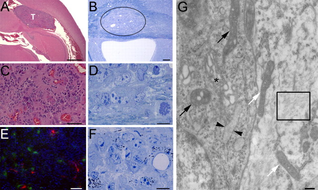
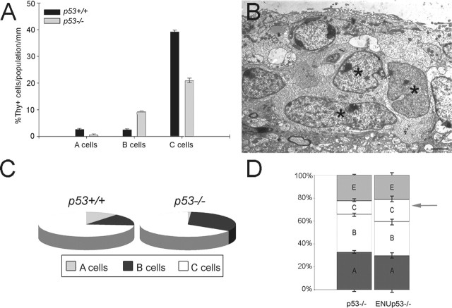
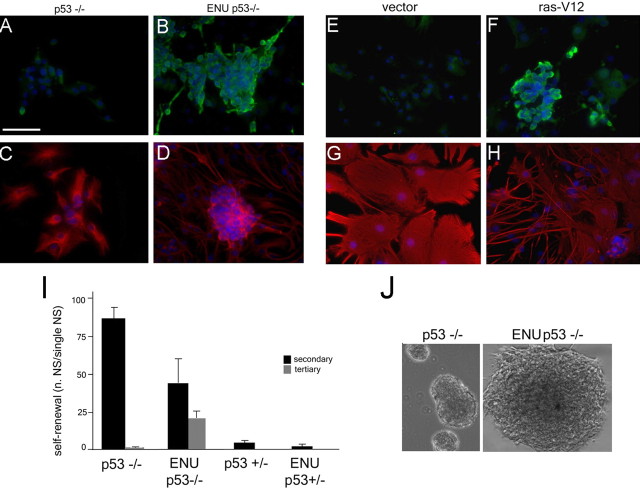
The role of multipotential progenitors and neural stem cells in the adult subventricular zone (SVZ) as cell-of-origin of glioblastoma has been suggested by studies on human tumors and transgenic mice. However, it is still unknown whether glial tumors are generated by all of the heterogeneous SVZ cell types or only by specific subpopulations of cells. It has been proposed that transformation could result from lack of apoptosis and increased self-renewal, but the definition of the properties leading to neoplastic transformation of SVZ cells are still elusive. This study addresses these questions in mice carrying the deletion of p53, a tumor-suppressor gene expressed in the SVZ. We show here that, although loss of p53 by itself is not sufficient for tumor formation, it provides a proliferative advantage to the slow- and fast-proliferating subventricular zone (SVZ) populations associated with their rapid differentiation. This results in areas of increased cell density that are distributed along the walls of the lateral ventricles and often associated with increased p53-independent apoptosis. Transformation occurs when loss of p53 is associated with a mutagenic stimulus and is characterized by dramatic changes in the properties of the quiescent adult SVZ cells, including enhanced self-renewal, recruitment to the fast-proliferating compartment, and impaired differentiation. Together, these findings provide a cellular mechanism for how the slow-proliferating SVZ cells can give rise to glial tumors in the adult brain.


Figures

Figure 1.
Figure 1.
Loss of p53 increases the number of adult neural stem cells and neuroblasts. A, RT-PCR (top) for the detection of p53 mRNA in adult mouse SVZ and Western blot (bottom) for the detection of p53 protein in SVZ protein extracts. Note the weak band detected in the SVZ extracts compared with HeLa cells. Actin was used as loading control. B, Detail of the lateral wall of the ventricle in irradiated (irrad) and not irradiated (not irrad) C57BL/6 wild-type mice. Note that p53 immunoreactivity after irradiation was restricted to cells of the SVZ, whereas the ependymal cells lining the lumen of the ventricles were not immunoreactive. Scale bar, 10 μm. C, Semithin sections of the SVZ in p53+/+ and p53−/− mice stained with toluidine blue showing the thickening of the cellular layer. Scale bar, 25 μm. D, Bar graph of the average number of SVZ cells identified per unit length in p53+/+ (black bars; n = 5) and p53−/− (gray bars; n = 5) mice. *p < 0.25; **p < 0.01.
Figure 2.
Figure 2.
Topographic map showing the heterogeneous distribution of cell types in the wall of the lateral ventricles of p53−/− mice compared with p53+/+. Topographic map of the SVZ determined by electron microscopy analysis and reconstruction of sequential sections. Each cell type is represented by a dot (red, A cells; blue, B cells; green, C cells; v, lateral ventricle); the ependymal cells lining the ventricular lumen are not shown. The boxed area corresponds to the adjacent enlarged semithin sections of the anterolateral superior corner and of the inferior border of the SVZ of p53+/+ and p53−/− mice stained with toluidine blue (v, lateral ventricle). Although some individual variability was observed in the length of the SVZ among different mice, no statistically significant difference was detected between the two genotypes.
Figure 3.
Figure 3.
Loss of p53 confers a proliferative advantage to fast-proliferating population and to quiescent type B cells. A, Immunoreactive BrdU+ cells detected in the anterior horn of the SVZ of p53+/+ and p53−/− littermates after a 1 h pulse labeling. Scale bar, 120 μm. B, Bar graph representing the number of BrdU+ cells per unit area in p53+/+ (black bar) and p53−/− (gray bar) mice. The asterisks indicate statistical significance, *p < 0.05. C, Toluidine blue-stained semithin sections of proliferating cells (arrows) in the lateral wall of the ventricles after [3H]Thy incorporation and autoradiography (v, ventricular lumen). Scale bar, 10 μm. D, Bar graph representing the total number of [3H]Thy cells per surface area in the SVZ of p53+/+ (black bar) and p53−/− (gray bar) mice. The asterisks indicate statistically significant differences between the two groups, *p < 0.05. E, The bar graphs indicate the percentage of cells in S-phase in each subpopulation in the SVZ of p53+/+ (black bar) and p53−/− (gray bar) mice identified by electron microscopy. F, Long-term retention of BrdU labeling of neural stem cells in the SVZ. Note the presence of a single cell in p53+/+ and several cells in the p53−/− littermates (v, ventricular lumen). Scale bar, 25 μm.
Figure 4.
Figure 4.
Effect of p53 loss-of-function on neurosphere-forming SVZ cells. A, Representative bright-field picture of secondary neurospheres generated from p53+/+ and p53−/− mice. Note the larger size of the spheres in the p53−/− cultures. B, Quantification of self-renewal in p53+/+ and p53−/− high- and low-density cultures. *p < 0.05. C, The amplification rate, measuring the total number of viable cells divided by the number of plated cells, was statistically higher in p53−/− mice; ***p < 0.001. D, Self-renewal rate in p53+/+ and p53flox/flox low-density cultures, either uninfected or infected with adenoviral vectors expressing Cre. The Cre-infected p53flox/flox cultures formed a larger number of neurospheres than uninfected or Cre-infected p53+/+. **p < 0.005. E, The number of viable cells in p53flox/flox cultures infected with adenoviral vectors expressing Cre recombinase was significantly higher than the number detected in Cre-infected p53+/+ cells; ***p < 0.001. Note the remarkable similarity of the results obtained in the Cre-infected p53flox/flox and p53−/− cultures. WT, Wild type.
Figure 5.
Figure 5.
Loss of p53 increases the number of neuroblasts both in vivo and in vitro. A, Immunofluorescence of the anterolateral superior corner of the SVZ stained with antibodies against PSA-NCAM (red) to identify the presence of neuroblastic migratory A cells. Note the increased immunoreactivity in p53−/− mice. Scale bar, 250 μm. B, C, Immunocytochemistry of p53+/+ and p53−/− SVZ cells differentiated for 7 d in vitro and stained with antibodies against the neuronal marker Tuj1 (B) and the oligodendrocyte marker O4 (C). Note the increased number of TuJ1+ and O4+ cells in p53−/− compared with the p53+/+ cultures. Scale bar, 180 μm. D, Quantification of the results illustrated in B. *p < 0.05; **p < 0.01). E, Immunocytochemistry of p53+/+ and p53flox/flox cultures, uninfected or infected with adenoviral vectors expressing Cre recombinase and then differentiated for 7 d in vitro. Cultures were stained with antibodies against the neuronal marker Tuj1 (red) to detect neuroblasts and against Cre (green) to detect infected cells. Note the increased number of TuJ1+ cells in the Cre-infected p53flox/flox cultures compared with p53+/+.
Figure 6.
Figure 6.
Spontaneous apoptosis is increased in the hyperplastic areas of the p53−/− SVZ. A, TUNEL assay in vivo in the SVZ of p53−/− mice, showing the presence of several apoptotic cells (green nuclei) within areas of localized hyperplasia (blue indicates DAPI as nuclear counterstain). B, TUNEL assay in vitro in cells dissociated from p53+/+ and p53−/− secondary neurospheres. Apoptotic nuclei are identified by arrows (TUNEL+, green; DAPI, blue as nuclear counterstain). Scale bar, 180 μm.
Figure 7.
Figure 7.
Glioblastoma-like tumors are detected in periventricular locations in adult p53−/− mice that have been exposed to the mutagen ENU. A, Micrograph of a sagittal section stained with hematoxylin–eosin showing a large tumoral mass (T) in periventricular location. Scale bar, 500 μm. B, Semithin section stained with toluidine blue showing a tumoral mass invading the corpus callosum and displacing the myelinated axons (circle). Scale bar, 75 μm. C, High-magnification view of the tumor, showing anaplastic cells, extensive microvascularization, and hemorrhages typical of glioblastomas. Scale bar, 50 μm. D, Very pleomorphic and anaplastic cells with dark cytoplasm, giant and invaginated nuclei with numerous chromatin clusters, and enormous nucleoli. Scale bar, 10 μm. E, Immunohistochemical analysis of the tumoral mass showing the presence of nestin+ (green) and GFAP+ (red) cells. DAPI+ (blue) was used as nuclear counterstain. Scale bar, 200 μm. F, Increased thymidine incorporation (after 1 h injection) is observed in cells within the tumor and more prominently at the periphery. Scale bar, 10 μm. G, Ultrastructural detail of a tumoral cell (left) contacting a nontumoral astrocyte (right). Note the moderately electron-dense cytoplasm of the tumoral cell (left) compared with the normal astrocyte and the lack of intermediate filaments (rectangle). The tumoral cell is also characterized by the presence of vacuoles in the mitochondria (black arrows) compared with normal cells (white arrows), enlarged Golgi apparatus (black asterisk) compared with control (white asterisk), and abundant and dilated endoplasmic reticulum (arrowheads). Scale bar, 200 nm.
Figure 8.
Figure 8.
Prenatal exposure of p53−/− mice to ENU induces the formation of periventricular glioblastoma-like tumors and is associated with the recruitment of the quiescent type B cells to the fast-proliferating compartment. A, Bar graph representing the number of [3H]Thy+ cells per unit area in each population of SVZ cells in untreated (gray) and ENU-treated (black) p53−/− mice. These data indicate that, during ENU treatment, the relative proportions of proliferating C and A cells are decreased, whereas the proportion of proliferating B cells is increased. B, Ultrastructural appearance of activated C cells (asterisks) in ENU-treated p53−/− mice. Note the presence of two C cells with giant nuclei and dispersed chromatin (left) and one cell with condensed chromatin (right, arrow), likely representing a B cell in transition. Ultrastructurally, C cells are characterized by the presence of large nuclei with invaginations and several nucleoli, a cytoplasm with abundant ribosomes, few mitochondria, endoplasmic reticulum cisternae, and a small Golgi apparatus. Scale bar, 2 μm. Although in physiological conditions the C cells are found in association with A cells, in ENU-treated p53−/− mice the C cells do not give rise to A cells but continue to divide and form these aggregates. C, The pies indicate the relative contribution of each subpopulation of SVZ cells to the total number of proliferating [3H]Thy+ within after a 1 h labeling period. Note the greater proportion of B cells in S-phase in the ENU-treated p53−/− mice. D, Relative shift of population dynamics before transformation. There is an accumulation of cells with intermediate feature between type B and C cells, with an expansion of the B/C compartment (arrow) at the expenses of the more differentiated neuroblasts.
Figure 9.
Figure 9.
SVZ cells cultured from ENU-treated p53−/− mice show enhanced self-renewal and impaired differentiation properties. A, Clonal self-renewal in mice of the indicated genotype that were exposed to the carcinogen ENU during the prenatal life. The bar graph indicates the average number of spheres generated by the dissociation of a single sphere after the second (black) or third (gray) passage. B, Note the large size of the ENU-treated p53−/− sphere even after repeated passaging. C–F, Nestin (green in C, E), GFAP (red in D, F), and DAPI (blue in C–F) immunofluorescence indicating the persistence of large aggregates of immature nestin+ cells and abnormal GFAP+ cells in cultures from ENU-treated p53−/− mice (F). G–J, Nestin (green in G–I) and GFAP (red in H–J) immunofluorescence reveals similar aggregates of nestin+ cells (I) and astrocytes displaying aberrant morphology (red in J) only in p53−/− cells expressing constitutively active Ras (Ras*). Scale bar, 75 μm.

Similar articles

Cited by

References

    1. Alvarez-Buylla A, Lim DA (2004). For the long run: maintaining germinal niches in the adult brain. Neuron 41:683–686. - PubMed
    1. Bachoo RM, Maher EA, Ligon KL, Sharpless NE, Chan SS, You MJ, Tang Y, DeFrances J, Stover E, Weissleder R, Rowitch DH, Louis DN, DePinho RA (2002). Epidermal growth factor receptor and Ink4a/Arf: convergent mechanisms governing terminal differentiation and transformation along the neural stem cell to astrocyte axis. Cancer Cell 1:269–277. - PubMed
    1. Berger F, Gay E, Pelletier L, Tropel P, Wion D (2004). Development of gliomas: potential role of asymmetrical cell division of neural stem cells. Lancet Oncol 5:511–514. - PubMed
    1. Bouvier C, Bartoli C, Aguirre-Cruz L, Virard I, Colin C, Fernandez C, Gouvernet J, Figarella-Branger D (2003). Shared oligodendrocyte lineage gene expression in gliomas and oligodendrocyte progenitor cells. J Neurosurg 99:344–350. - PubMed
    1. Chow BM, Li YQ, Wong CS (2000). Radiation-induced apoptosis in the adult central nervous system is p53-dependent. Cell Death Differ 7:712–720. - PubMed

Publication types

MeSH terms

Substances