. 2006 Jan 25;26(4):1154-63.
doi: 10.1523/JNEUROSCI.4424-05.2006.

Dendrites contain a spacing pattern

Affiliations

Dendrites contain a spacing pattern

Aaron B Taylor et al. J Neurosci. .

Abstract

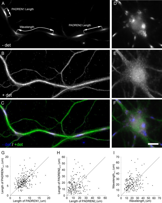
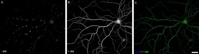
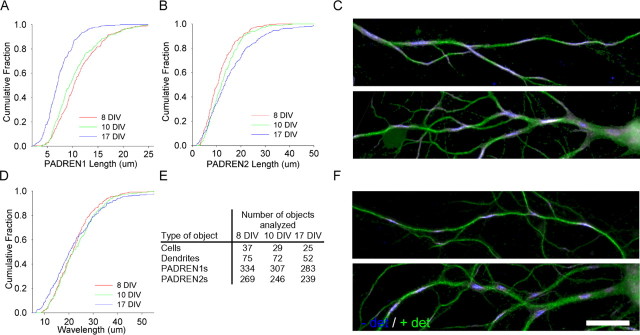
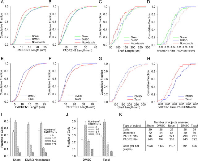
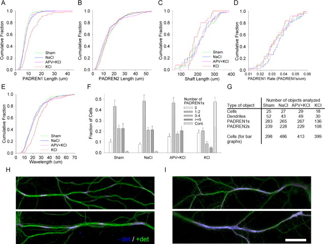
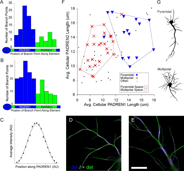
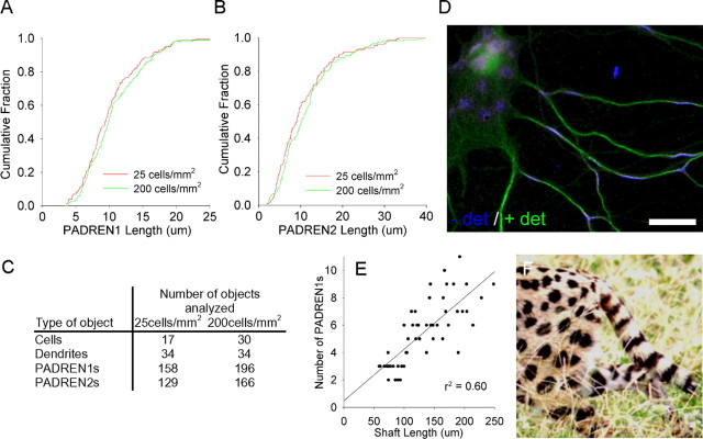
The distinctive branching patterns of dendritic arbors are essential for neuronal information processing. The final shape of an arbor is the result of intrinsic and extrinsic factors. However, the cellular mechanisms that underlie branch patterning are unknown. In many biological systems, locally acting factors are intrinsically organized into spacing patterns that guide patterned morphogenesis. Here, we show that neurons contain two types of periodic and regular elements (PADREN1s and PADREN2s) that are arranged into a spacing pattern. The wavelength of the pattern is approximately 20 microm. Dendritic branches occur preferentially within PADREN1s, and specific PADREN lengths correspond to specific arbor types. The lengths of the PADRENs also change over time and can be modified by activity. However, PADRENs are intrinsically organized, possibly by a reaction-diffusion process. PADRENs reveal a previously unrecognized level of neuronal organization that may provide insight into how the distinct branching patterns of the dendrites are intrinsically organized.


Figures

Figure 1.
Figure 1.
PADRENs form a spacing pattern. PADREN stripes (A) in the dendrite and spots (D) in the soma of a 17 DIV hippocampal neuron. Signal represents immunostaining before application of detergent (− det) using the mouse 5F9 anti-MAP2 antibody. Leaders in A demarcate a PADREN1 length, PADREN2 length, and a wavelength. B, E, Signal represents immunostaining after application of detergent (+ det) using rabbit polyclonal anti-MAP2 antibodies. C, F, Merged signals. Scale bar, 10 μm. G–I, Scatter plots of adjacent (ith + 1 vs ith) PADREN1 lengths (G), PADREN2 lengths (H), and wavelengths (I) in 17 DIV cells show that adjacent lengths are similar. The line Lengthi + 1 = Lengthi is illustrated for comparison.
Figure 2.
Figure 2.
PADRENs form a spacing pattern. PADRENs in a 17 DIV hippocampal neuron. A, Immunostaining before application of detergent (− det) using the mouse 5F9 anti-MAP2 antibody. B, Immunostaining after application of detergent (+ det) using rabbit polyclonal anti-MAP2 antibodies. C, Merge. Scale bar, 30 μm.
Figure 3.
Figure 3.
PADREN lengths change during development. PADREN lengths change with cell age from 8 to 17 DIV (PADREN1 lengths, p < 1 × 10⁻¹⁰; PADREN2 lengths, p = 2.5 × 10⁻⁵; Kruskal–Wallis test). Cumulative distributions showing PADREN1 lengths decrease across development (A), whereas PADREN2 lengths increase (B) (PADREN1 lengths, 8 vs 10 DIV, p = 0.14; 10 vs 17 DIV, p = 5 × 10⁻⁹; PADREN2 lengths, 8 vs 10 DIV, p = 0.057; 10 vs 17 DIV, p = 0.001; Tukey’s honestly significant difference post hoc multiple comparisons test). D, Remarkably, wavelength is conserved (p = 0.24, Kruskal–Wallis test). E, The table shows the type (left column) and number (right columns) of objects analyzed at each time point to produce the data in A, B, and D. Cell density decreased 20% during this time interval (data not shown). Examples of PADRENs in 10 DIV (C) and 17 DIV (F) hippocampal neurons. Scale bar, 20 μm.
Figure 4.
Figure 4.
PADRENs scale with dendritic lengths. Shaft lengths and PADREN lengths scale correspondingly in 10 DIV hippocampal neurons. Cumulative distributions showing that nocodazole (15 μg/ml, 2 h) causes PADREN1 lengths (A), PADREN2 lengths (B), and dendritic shaft lengths (C) to significantly scale down (nocodazole vs DMSO, PADREN1 lengths, p = 2.9 × 10⁻⁶; PADREN2 lengths, p = 1.4 × 10⁻⁵; shaft lengths, p = 0.012). There is no evidence that the number of PADRENs per branch changed, because the PADREN1 rate (number of PADREN1s per length of shaft) increased (p = 0.011), as expected if shafts became shorter but contained the same number of PADRENs (D). Conversely, in the presence of taxol (500 nm, 4 h), PADREN1 lengths (E), PADREN2 lengths (F), and dendritic shaft lengths (G) significantly scale up [taxol vs DMSO, PADREN1 lengths, p = 5.6 × 10⁻⁷; PADREN2 lengths, p = 0.002; shaft lengths, p = 0.037 (an additional 35 DMSO-treated shaft lengths and 30 taxol-treated shaft lengths from a separate experiment were added to achieve a p < 0.05 increase in shaft length)]. Again, there is no evidence that the number of PADRENs changed, because the PADREN1 rate decreased significantly (p = 0.006), as expected if the number of PADRENs remained constant as the lengths of the shafts increased (H). I, J, Neither treatment changed the fraction of cells with at least one dendrite containing different numbers of PADREN1s or changed cell density (data not shown). Graph bars represent average fraction of cells per 40× field in each “Number of PADREN1s” category; error bars represent SEM. K, A table showing the type (left column) and number of objects analyzed (right columns) under each condition to produce the data shown in A–J.
Figure 5.
Figure 5.
PADREN1 lengths are modified by activity. A, A cumulative distribution showing that applying KCl (20 mm, 15 min) increases the length of PADREN1s compared with cultures treated with APV (100 μm, 1 h pretreatment) and then KCl (p < 1 × 10⁻¹⁰). The block with APV was complete because the APV plus KCl group was similar to the NaCl control (p = 0.8). We did not detect a change in PADREN2 length (B), and measured wavelength increased (E) (p = 3.25 × 10⁻⁴). Shaft lengths (C) were unaffected (KCl vs NaCl; p = 0.11), as were segmentation rates (D). F, A bar graph showing that KCl also changes the fraction of cells containing different numbers of PADREN1s or having continuous immunostaining. The graph bars represent average fraction of cells per 40× field in each category; error bars represent SEM. G, A table showing the type (left column) and number (right columns) of objects analyzed under each condition to produce the data shown in A–F. Examples of PADRENs in control (H) and KCl-treated (I) dendrites show that PADREN1s lengthen (I, top) and become continuous (I, bottom) in response to KCl. Scale bar, 20 μm.
Figure 6.
Figure 6.
Dendritic branch points preferentially associate with PADREN1s, and multipolar and pyramidal cells have distinct PADREN lengths. A, B, Frequency histograms showing the relative position of branch points along PADREN1s (blue) and PADREN2s (green) at 8 DIV (A) and 10 DIV (B). Relative position was computed as the distance from the proximal PADREN edge to the branch point divided by the length of the PADREN containing the branch point. “Soma” icons confer proximal–distal orientation to shaft icons. Branch points significantly associate with PADREN1s (p = 1.80 × 10⁻⁶, 8 DIV; p = 4.07 × 10⁻⁶, 10 DIV; custom test; for details, see Materials and Methods). Branch positions form a bell-shaped distribution along PADREN1s, which mirrors a Gaussian distribution of immunostaining intensity along PADREN1s (C). Points represent average immunostaining as a function of relative position after standardization and normalization from six randomly selected PADREN1s that did not contain branch points. D, E, Examples of the association between PADREN1s and branch points in 10 DIV neurons. Scale bar, 20 μm. F, Scatter plot of average cellular PADREN2 length versus average cellular PADREN1 length for each of 78 10 DIV cells. Point style indicates arbor type classification (pyramidal, multipolar, or other plotted as triangles, crosses, and dots, respectively). The shafts of pyramidal cells were approximately twice as long as the shafts of multipolar cells (pyramidal, 202 ± 14 μm; multipolar, 110 ± 4 μm; p = 1.75 × 10⁻⁸, t test). Showing PADREN dimensions and arbor types correspond, convex polygons enclosing pyramidal or multipolar cells delineate distinct PADREN dimension spaces. Both PADREN1s (p < 1 × 10⁻¹⁰) and PADREN2s (p = 0.006) are longer in pyramidal cells than multipolar cells. The chance of uniformly distributed pyramidal and multipolar cell types falling into the PADREN space areas they occupy is p ≪ 0.005 in both cases (χ2 test, df = 1). Additionally, average PADREN1 lengths and average PADREN2 lengths are correlated (β1 = 0.50; p1 = 0) = 2 × 10⁻⁶; simple linear regression; line not shown). G, Examples of arbor morphologies classified as pyramidal and multipolar. − det, Before the addition of detergent; + det, after the addition of detergent; AU, arbitrary units.
Figure 7.
Figure 7.
PADRENs develop autonomously and could be organized by a reaction-diffusion process. Cumulative distributions showing that changing the plating density eightfold across different wells of the same plate does not effect the length of PADREN1s (A) (p = 0.17) or PADREN2s (B) (p = 0.17) at 10 DIV. C, The table shows the type and number of objects analyzed under each condition. D, A 17 DIV hippocampal neuron with a particularly flat cell body illustrates the geometry-dependent transition of PADRENs from stripes in the narrow cylindrical dendrites to spots in the locally planar cell body. Scale bar, 20 μm. Such a transition is also observed in the coat markings of many animals, including the serval (F), and is consistent with a reaction-diffusion processes (serval photograph courtesy of Dr. Shonali Laha, Florida International University, Miami, FL). E, Further consistent with a reaction-diffusion process, a scatter plot showing a linear correlation between shaft length and the number of PADREN1s per shaft in 10 DIV hippocampal neurons.

References

    1. Agarwala S, Sanders TA, Ragsdale CW (2001). Sonic hedgehog control of size and shape in midbrain pattern formation. Science 291:2147–2150. - PubMed
    1. Agmon-Snir H, Carr CE, Rinzel J (1998). The role of dendrites in auditory coincidence detection. Nature 393:268–272. - PubMed
    1. Aizawa H, Hu SC, Bobb K, Balakrishnan K, Ince G, Gurevich I, Cowan M, Ghosh A (2004). Dendrite development regulated by CREST, a calcium-regulated transcriptional activator. Science 303:197–202. - PubMed
    1. Akum BF, Chen M, Gunderson SI, Riefler GM, Scerri-Hansen MM, Firestein BL (2004). Cypin regulates dendrite patterning in hippocampal neurons by promoting microtubule assembly. Nat Neurosci 7:145–152. - PubMed
    1. Ashford JW, Soultanian NS, Zhang SX, Geddes JW (1998). Neuropil threads are collinear with MAP2 immunostaining in neuronal dendrites of Alzheimer brain. J Neuropathol Exp Neurol 57:972–978. - PubMed

Publication types

MeSH terms

LinkOut - more resources