. 2006 Jan 25;26(4):1164-74.
doi: 10.1523/JNEUROSCI.3116-05.2006.

Transition from reversible to persistent binding of CaMKII to postsynaptic sites and NR2B

Affiliations

Transition from reversible to persistent binding of CaMKII to postsynaptic sites and NR2B

K Ulrich Bayer et al. J Neurosci. .

Abstract

Changes in protein-protein interactions and activity states have been proposed to underlie persistent synaptic remodeling that is induced by transient stimuli. Here, we show an unusual stimulus-dependent transition from a short-lived to long-lasting binding between a synaptic receptor and its transducer. Both molecules, the NMDA receptor subunit NR2B and Ca2+/calmodulin (CaM)-dependent protein kinase II (CaMKII), are strongly implicated in mediating synaptic plasticity. We show that CaMKII reversibly translocates to synaptic sites in response to brief stimuli, but its resident time at the synapse increases after longer stimulation. Thus, CaMKII localization reflects temporal patterns of synaptic stimulation. We have identified two surface regions of CaMKII involved in short-lived and long-term interactions with NR2B. Our results support an initial reversible and Ca2+/CaM-dependent binding at the substrate-binding site ("S-site"). On longer stimulation, a persistent interaction is formed at the T286-binding site ("T-site"), thereby keeping the autoregulatory domain displaced and enabling Ca2+/CaM-independent kinase activity. Such dual modes of interaction were observed in vitro and in HEK cells. In hippocampal neurons, short-term stimulation initiates a reversible translocation, but an active history of stimulation beyond some threshold produces a persistent synaptic localization of CaMKII. This activity-dependent incorporation of CaMKII into postsynaptic sites may play a role in maturation and plasticity of synapses.


Figures

Figure 1.
Figure 1.
Spontaneous postsynaptic CaMKIIα localization in hippocampal neurons (12–14 DIV). A, B, Spontaneous synaptic enrichment of CaMKIIα depends on NMDAR activity. Neurons expressing GFP-CaMKIIα were incubated without (A; W/O) or with (B) 100 μm AP-5 for 16 h, fixed, and immunostained for PSD95. Boxes indicate cropped area on the right. Arrowheads indicate synaptic sites. Colocalization was more pronounced in untreated neurons. Scale bars: overview, 10 μm; cropped, 5 μm. C, Mean percentage (±SEM; n = 3) of neurons (78–93 per n) exhibiting synaptic enrichment of GFP-CaMKIIα (neurons with localization score of “+” or more; see Materials and Methods). Neurons were transfected with wild-type (wt) or T305/306D GFP-CaMKIIα. The asterisk indicates significantly different from T305/306D (*p < 0.01; t test).
Figure 2.
Figure 2.
Stimulus-induced transition from reversible to persistent synaptic CaMKII localization. A, Time lapse images of GFP-CaMKIIα in a neuron preincubated with AP-5. Arrowheads point to examples of synapses in which GFP-CaMKIIα accumulated during the 1 min glutamate/glycine stimulation (stim) and persisted after the 30 min wash. Scale bars, 10 μm. B, Mean (±SEM) postsynaptic localization index of wild-type (wt) and T286A GFP-CaMKIIα in neurons treated as in A. The initial dissociation curves (inset) for both wild type and T286A differed significantly from pooled data (p < 0.002; F test). C, Effect of stimulation time on the poststimulation plateau of postsynaptic localization index of CaMKII. Mean (±SEM; n = 7–9) plateaus were determined between 15 and 30 min of washout. The asterisk indicates wild type and T286A differed significantly at 60 and 120 s from their 30 s time point (*p < 0.005; t test). Persistent plateaus of wt and T286A differed significantly from pooled data only at 120 s (p < 0.005; F test).
Figure 3.
Figure 3.
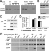
Ca2+-dependent and -independent states of CaMKIIα interaction with NR2B in vitro and in HEK cells. A, B, CaMKIIα was allowed to bind to immobilized GST-NR2B-C for various times, as indicated, at room temperature in the presence of Ca2+/CaM. Then, the complexes were washed either with Ca2+/CaM or EGTA. A, Bound CaMKIIα is detected by immunoblotting of eluted protein. B, CaMKIIα binding under the different conditions was compared by densitometry using NIH Image (means ± SEM; n = 6). The asterisks indicate wash conditions that yielded significantly different CaMKII binding after 0.5 min (p < 0.005; t test) and 1.5 min (p < 0.05; t test). C, Localization of GFP-CaMKIIα-T286A in living HEK cells cotransfected with NR2B changes after stimulation (stim.) with Ca2+/ionomycin. Localization before and after two rounds of stimulation with subsequent EGTA washes is shown; stimulation times (2 and 6 min) and wash times (10 and 45 min) are indicated. Note the appearance of punctated fluorescence after the stimuli, which persisted after longer (6 min) stimulation.
Figure 4.
Figure 4.
All CaMKII isoforms can bind to NR2B in vitro, but four mutants do not. A, B, Binding of CaMKII isoforms (A) and point mutants to immobilized GST-NR2B-C (B) was tested in vitro. Ca2+/CaM was present in the binding reaction but not in the washes. Bound CaMKII is visualized by CaM overlay (A) or immunoblotting (B) of the eluted protein. The bottom panels show half of the amount of kinase used in the binding assays. wt, Wild type. C, Three views of a CaMKII kinase domain model (Yang and Schulman, 1999). Amino acids: aqua, differ between isoforms; green, mutations that did not affect NR2B binding; yellow, mutations that impaired NR2B binding.
Figure 5.
Figure 5.
Impaired translocation of CaMKII mutants to NR2B in HEK cells. GFP-CaMKII isoforms or mutants were coexpressed with or without NR2B in HEK cells, and kinase localization before and after an ionomycin-induced Ca2+ stimulus was monitored. Representative cells are shown. The number of cells with translocating kinase per number of cells monitored (32–85) is indicated for each condition in the lower right corner of the corresponding panel. A, CaMKIIα and β wild-type translocate in an NR2B-dependent manner. B, The four CaMKIIα mutants with impaired NR2B-C-binding in vitro show impaired translocation in HEK cells coexpressing NR2B. Pictures were taken 6 min after stimulation unless indicated otherwise. Only the W237K mutant showed any NR2B-dependent translocation.
Figure 6.
Figure 6.
CaMKIIα mutants with impaired NR2B binding show reduced postsynaptic localization. Relative frequency plot of postsynaptic localization of GFP-CaMKIIα wild type (wt) and mutants after a 1 min glutamate/glycine stimulation and an immediate fixation (neurons cultured as in Fig. 1b). 1–326 is a monomeric CaMKIIα mutant. The right panels show examples of the dendrite categories used for scores (− to +++). Every transfected neuron (14 DIV; 40–80 per coverslip; n = 2) was scored for each GFP-CaMKIIα construct tested. Scale bar, 5 μm.
Figure 7.
Figure 7.
The NR2B-binding surface on CaMKII is involved in autoregulation. A, The residues implicated in the interaction with the autoregulatory region (shown as trace) are marked in this model of a kinase domain. Orange, Substrate binding site (S-site); yellow, T286 interacting site (T-site); boxed area, mutations that impaired NR2B binding. B, Alignment of the CaMKIIα autoregulatory region and the binding region on NR2B. Proposed T- and S-site interactions are marked in yellow and orange, respectively. The autoregulatory region of CaMKII interacts with the kinase domain of the same subunit as indicated in the top panel or with a neighboring subunit during T286 autophosphorylation as indicated in the bottom panel. Possible NR2B-specific additional S-site interactions of CaMKII I205 are indicated by dashed orange lines. C, Models for biphasic interaction of a CaMKII kinase domain with NR2B. i, Basal state; ii, Ca2+/CaM allows S-site access; iii, initial S-site binding of NR2B is required for T-site access (but can be circumvented by peptides occupying the S-site); iv, dissociation-reassociation provides stimulus time-dependent step; v, T-site binding involves more amino acids and is persistent.
Figure 8.
Figure 8.
Effects of T- and S-site binding peptides on CaMKIIα interaction with NR2B. A, CaMKIIα (0.2 μm) was incubated with different peptides (pept.) before the binding reaction to immobilized NR2B-C. Bound kinase was detected by immunoblotting the eluted protein. Syntide (syn) binds to the kinase S-site, whereas AC3 can interact with both the S-site and the T-site. N2B-l and N2B-s are derived from amino acids 1259–1310 and 1289–1310 of NR2B, respectively. B, CaMKII was allowed to bind to immobilized NR2B-C; then, different peptides were added and incubated for 1 h. Essentially, all of the kinase remained bound (top), and no kinase was detected in the supernatant (bottom). C, N2B-s (NR2B1289–1310) but not syntide affects CaMKII-NR2B interaction at high concentration. D, Autonomous kinase activity (means ± SEM) of CaMKIIα holoenzymes (CaMKII wt) and monomers (1–316) are affected similarly by the NR2B-derived peptides. Kinase/peptide interaction was enabled by Ca2+/CaM before the addition of EGTA. Autonomy is expressed as a percentage of the activity stimulated by Ca2+/CaM (in presence of peptide). E, Time course of CaMKIIα binding to NR2B-C. Kinase, peptides, and Ca2+/CaM were preincubated for 8 min. Bottom, CaM was dissociated from the kinase by EGTA before the binding reaction. Binding reactions were done at 4°C to slow down the kinetics.
Figure 9.
Figure 9.
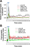
Effect of S- and T-site mutations on the transient and persistent postsynaptic translocation of CaMKIIα after a 1 min glutamate/glycine stimulus. A, Mean (±SEM) postsynaptic localization of wild type (wt) and mutant GFP-CaMKIIα relative to basal state (averaged over the first 2 min) measured before, during, and after stimulation (stim.) (sample images are shown in supplemental Fig. 2, available at www.jneurosci.org as supplemental material). Statistical differences (t test) for the peak values compared with wt (0.167 ± 0.051) are p < 0.024 (I205K, 0.049 ± 0.003), p < 0.042 (E139R, 0.065 ± 0.014), and p < 0.056 (W237K, 0.071 ± 0.012). B, Mean (±SEM) postsynaptic localization index of wt and mutant GFP-CaMKIIα in neurons treated as in A. Data from A are rescaled from 0 (basal postsynaptic localization state, averaged over the first 2 min) to 1 (maximal postsynaptic localization) to compare the postsynaptic dissociation of the different mutants and wt CaMKII after the stimulus. The E139R mutant also exhibited a long-lasting plateau of postsynaptic enrichment after the stimulus. The statistical differences (t test) for the mean index from 15 to 30 min of wash compared with wt (0.218 ± 0.011) are p < 0.006 (W237K, 0.034 ± 0.003), p < 0.042 (I205K, 0.064 ± 0.004), and p > 0.1 (E139R, 0.195 ± 0.011).

References

    1. Barria AS, Malinow R (2004). NMDA receptor subunits and synaptic plasticity. Soc Neurosci Abstr 30:55–20.
    1. Barria A, Malinow R (2005). NMDA receptor subunit composition controls synaptic plasticity by regulating binding to CaMKII. Neuron 48:289–301. - PubMed
    1. Bayer KU, Schulman H (2001). Regulation of signal transduction by protein targeting: the case for CaMKII. Biochem Biophys Res Commun 289:917–923. - PubMed
    1. Bayer KU, Harbers K, Schulman H (1998). alphaKAP is an anchoring protein for a novel CaM kinase II isoform in skeletal muscle. EMBO J 17:5598–5605. - PMC - PubMed
    1. Bayer KU, De Koninck P, Leonard AS, Hell JW, Schulman H (2001). Interaction with the NMDA receptor locks CaMKII in an active conformation. Nature 411:801–805. - PubMed

Publication types

MeSH terms

LinkOut - more resources