. 2006 Jan 25;26(4):1179-89.
doi: 10.1523/JNEUROSCI.2618-05.2006.

Peripheral myelin protein 22 is in complex with alpha6beta4 integrin, and its absence alters the Schwann cell basal lamina

Affiliations

Peripheral myelin protein 22 is in complex with alpha6beta4 integrin, and its absence alters the Schwann cell basal lamina

Stephanie A Amici et al. J Neurosci. .

Abstract

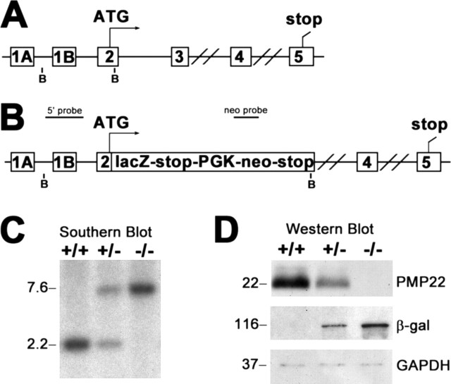
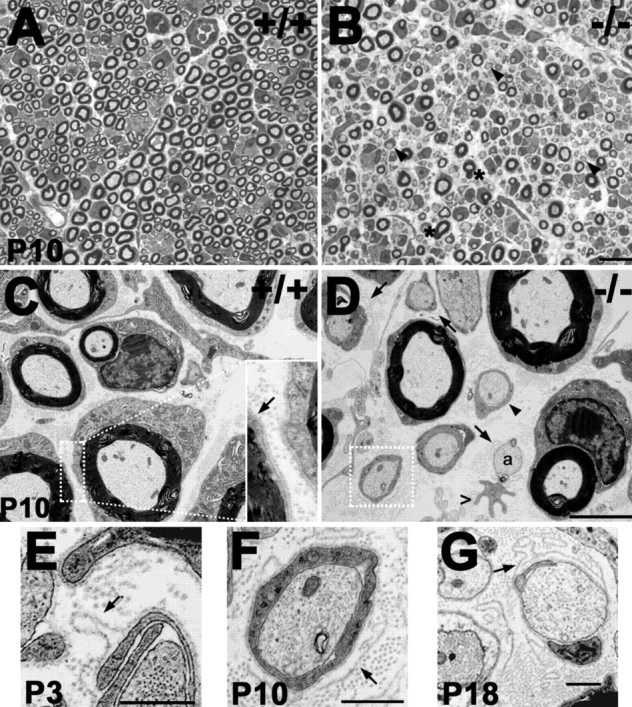
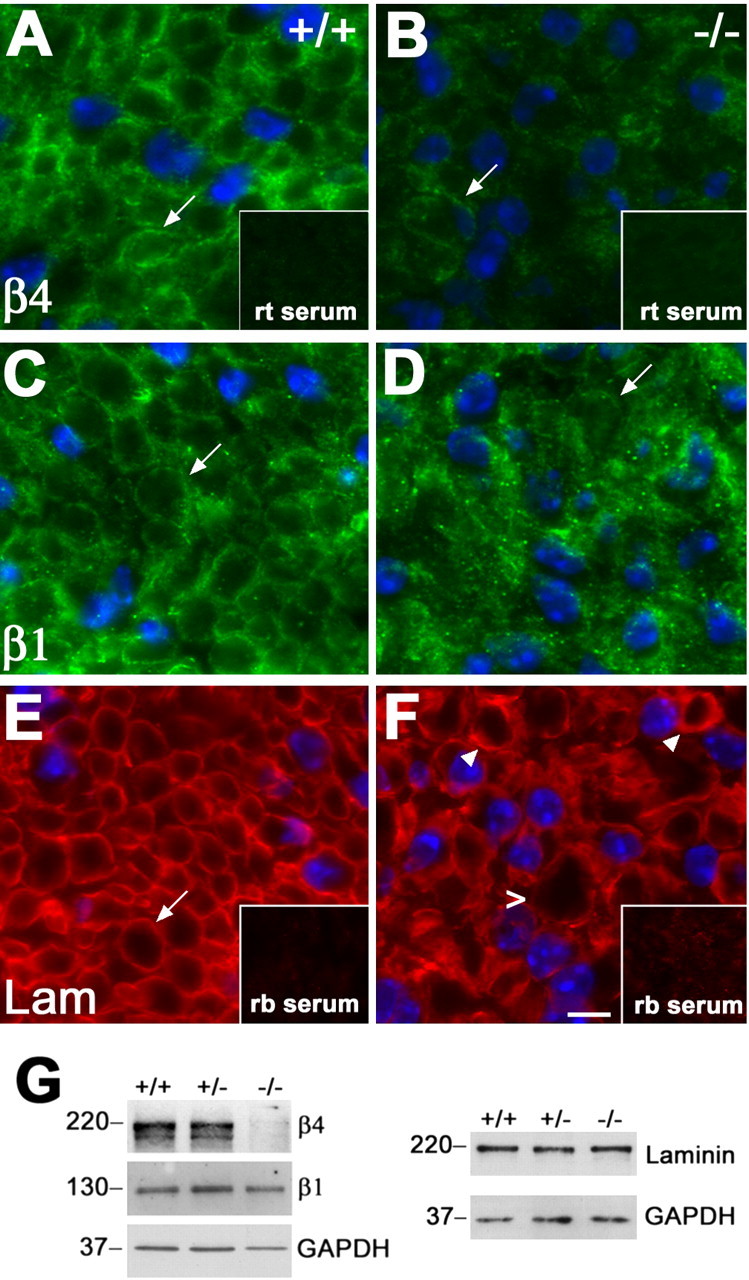
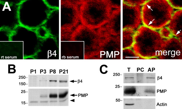
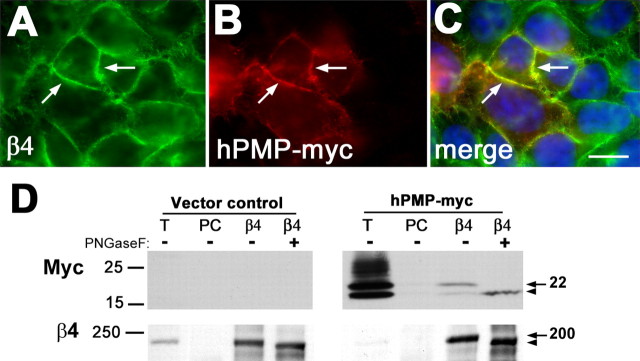
Peripheral myelin protein 22 (PMP22) is a tetraspan membrane glycoprotein, the misexpression of which is associated with hereditary demyelinating neuropathies. Myelinating Schwann cells (SCs) produce the highest levels of PMP22, yet the function of the protein in peripheral nerve biology is unresolved. To investigate the potential roles of PMP22, we engineered a novel knock-out (-/-) mouse line by replacing the first two coding exons of pmp22 with the lacZ reporter. PMP22-deficient mice show strong beta-galactosidase reactivity in peripheral nerves, cartilage, intestines, and lungs, whereas phenotypically they display the characteristics of tomaculous neuropathy. In the absence of PMP22, myelination of peripheral nerves is delayed, and numerous axon-SC profiles show loose basal lamina, suggesting altered interactions of the glial cells with the extracellular matrix. The levels of beta4 integrin, a molecule involved in the linkage between SCs and the basal lamina, are severely reduced in nerves of PMP22-deficient mice. During early stages of myelination, PMP22 and beta4 integrin are coexpressed at the cell surface and can be coimmunoprecipitated together with laminin and alpha6 integrin. In agreement, in clone A colonic carcinoma cells, epitope-tagged PMP22 forms a complex with beta4 integrin. Together, these data indicate that PMP22 is a binding partner in the integrin/laminin complex and is involved in mediating the interaction of SCs with the extracellular environment.


Figures

Figure 1.
Figure 1.
A, Schematic representation of the targeting strategy. The genomic structure of the mouse pmp22, showing the alternatively used exons 1A and 1B, which are preceded by promoters 1 and 2, respectively, is shown. The double bars between exons 3 and 4 and exons 4 and 5 indicate 16.6 and 7.0 kb intronic sequences, respectively. B, Diagram of the pmp22-lacZ construct. The first two coding exons of pmp22 (exons 2 and 3) were replaced with the lacZ reporter, followed by a strong transcription termination signal, the PGK-neo gene, and a second stop signal. BamHI restriction sites (B) and locations of the PMP22 (5′ probe) and neo probes are shown (A, B). C, Southern blot analysis of genomic DNA digested with BamHI from wild type (+/+), heterozygous (+/−), and homozygous PMP22 knock-out (−/−) littermates. The endogenous allele is detected as a 2.2 kb fragment, whereas the mutant allele yields a 7.6 kb fragment. D, Western blot analyses of sciatic nerve lysates (7.5 μg/lane) from adult +/+, +/−, and −/− mice with anti-PMP22 and anti-β-gal antibodies. Anti-GAPDH is shown as a loading control. Molecular mass in kilobases (C) and kilodaltons (D) is shown on the left.
Figure 2.
Figure 2.
Widespread β-gal expression in the E15 mouse embryo. A, The β-gal reporter is expressed in a variety of neural and non-neural tissues on a cryosection of an E15 embryo. B, C, Higher-magnification images show reporter expression in the ventricular zones of the rhombencephalon (R) and mesencephalon (M) (10×; B), DRG (8×; C, arrow), skeletal muscle (C, asterisk), and cartilage of the vertebrae (C, arrowhead). D, β-Gal activity is prominent in the aortic arch of the heart (7×; arrow). E, F, High levels of the reporter are also present in the lungs (18×; E) and the intestines (9×; F). The sections are counterstained with neutral red.
Figure 3.
Figure 3.
Phenotype of adult PMP22-deficient mice. A–D, Distinct β-gal expression is evident in the ribs (A), aortic arch of the heart (B), brainstem deep nuclei (C, arrows), cranial nerve roots (C, arrowheads), and the peripheral roots of a spinal cord preparation (D). E, Behavioral phenotypes of 3-month-old littermates. Wild type (+/+) mice have splayed hindlimbs and relaxed forelimbs, whereas PMP22-deficient littermates (−/−) clasp their hindlimbs and forelimbs close to their body (top right) and display gait abnormalities (bottom). F, Teased nerve fibers from genotyped P21 littermates. Nerve fibers of PMP22−/− mice exhibit frequent tomacula (arrowhead). Magnification, 40×.
Figure 4.
Figure 4.
Delayed myelination and ultrastructural alterations in nerves of affected mice. A, B, Semithin (1 μm thickness) toluidine blue-stained cross sections from P10 wild-type (A; +/+) and PMP22-deficient (B; −/−) littermates. In contrast to the normal nerve (A), there is a delay in myelination in PMP22-deficient samples, because many promyelinating fibers are observed (B, arrowheads). Thickened myelin profiles are also present (B, asterisks). C, On ultrastuctural examination, nerves of P10 +/+ mice show tight basal lamina (arrow in inset; 3× magnification) around the SCs. D–G, In comparison, promyelinating SCs (arrowhead), loose basal lamina (arrows), bare axons (a), and irregular, hypertrophic SC processes (open arrowhead) are evident in the P10 −/− sample. The boxed area in D is magnified in F (3×). SC profiles with pockets of loose basal lamina are also seen in nerves from P3 (E) and P18 (G) PMP22−/− mice. Scale bars: A, B, 10 μm; C, D, 2 μm; C (inset), F, 0.67 μm; E, G, 0.5 μm.
Figure 5.
Figure 5.
Decreased levels of β4 integrin in nerves of PMP22-deficient mice. A–F, Cryosections of sciatic nerves from +/+ (A, C, E) and PMP22−/− (B, D, F) mice were immunostained with monoclonal rat (rt) anti-β4 integrin (A, B), anti-β1 integrin (C, D), or polyclonal rabbit (rb) anti-laminin (Lam) (E, F) antibodies. In nerves of P10 +/+ mice, β4 and β1 integrin are detected at the abaxonal SC surface (A, C, arrows). In comparison, abaxonal integrin-like staining is only discernable around a fraction of the fibers in the −/− samples (B, D, arrows). In addition, when the images were collected at the same exposure times, the level of β4-like immunoreactivity was reduced (compare A, B). Laminin was detected at the SC basal lamina in +/+ (E, arrow) and −/− (F) nerves, with thickened basal lamina (arrowheads) and a tomaculum (open arrowhead) marked in the affected sample (F). Nonspecific rat (A, B, inset) and rabbit (E, F, inset) sera serve as controls for staining specificity. Scale bar, 5 μm. G, Sciatic nerve lysates (20 μg/lane) from P10 +/+, +/−, and −/− mice were analyzed with polyclonal rabbit anti-β4 integrin and anti-laminin and monoclonal rat anti-β1 integrin antibodies. The blots were reprobed with monoclonal mouse anti-GAPDH antibody as a protein loading control. Molecular mass is in kilodaltons.
Figure 6.
Figure 6.
PMP22 is in a complex with α6β4 integrin and laminin. Sciatic nerve lysates (T lanes) from P21 +/+ mice were processed for IP, after preclearing (PC lanes) with nonspecific Igs of the appropriate isotype. Lysates were incubated with polyclonal rabbit anti-β4 integrin (A, left, C), anti-PMP22 (A, right), monoclonal rat anti-α6 integrin (B, left), or polyclonal rabbit anti-laminin (B, right) antibodies, and captured immunoprecipitates were probed for the indicated proteins (A–C) as designated at the right of each blot. On reprobes (marked with asterisks), after stripping the membranes, the anti-β4 integrin and anti-laminin antibodies did not work efficiently when 1 μg/lane total (T) nerve protein was analyzed. IP with anti-β4 integrin on nerve lysates of homozygous PMP22-deficient mice is shown as a negative control (C). Molecular mass is in kilodaltons. PMP, PMP22; Lam, laminin; βDys, β-dystroglycan.
Figure 7.
Figure 7.
Coexpression of PMP22 and integrins during myelination. A, Sciatic nerve cryosections from 3-month-old +/+ mice were labeled with monoclonal rat anti-β4 and polyclonal rabbit anti-PMP22 antibodies and examined by confocal microscopy. The merged single plane image reveals the partial colocalization (merge, yellow) of β4 integrin (green) and PMP22 (red). Scale bar, 5 μm. Nonspecific rat (rt) and rabbit (rb) sera (bottom insets) serve as controls for staining specificity. B, Entire protein lysates of P1, P3, P8, and P21 sciatic nerves (10 μg/lane) from +/+ mice were analyzed with anti-PMP22 and anti-β4 integrin antibodies. The arrows indicate the migration of β4 integrin at ∼200 kDa (top) and of PMP22 at ∼22 kDa (bottom), whereas the arrowhead marks a nonspecific immunoreactive band. C, Cell-surface biotinylation of myelinating DRG–SC cocultures identifies β4 integrin and PMP22 in the avidin pull down (AP), from which actin is excluded. Total lysate (T) and agarose bead preclear (PC) fractions are also shown. Molecular mass is in kilodaltons. PMP, PMP22.
Figure 8.
Figure 8.
PMP22 and β4 integrin are coimmunoprecipitated from clone A cells. A, B, Epitope (myc)-tagged hPMP22 was expressed in clone A cells, and samples were double immunolabeled with anti-β4 (A) and anti-myc (B) antibodies. C, As the merged image reveals, hPMP22 is targeted to the β4 integrin-positive plasma membrane of the cells (A–C, arrows). Scale bar, 10 μm. D, Vector control and hPMP-myc-expressing clone A cells were lysed and processed for IP with the indicated antibodies. The precipitates were subsequently probed for the marked proteins by Western blot. The arrows on the right indicate the overexpressed glycosylated PMP22 (∼22 kDa) and the endogenous β4 integrin (∼200 kDa). The arrowheads point at the position of the deglycosylated ∼18 and ∼190 kDa forms. Molecular mass is in kilodaltons. T, Total lysate; PC, preclear.

References

    1. Adlkofer K, Martini R, Aguzzi A, Zielasek J, Toyka KV, Suter U (1995). Hypermyelination and demyelinating peripheral neuropathy in Pmp22-deficient mice. Nat Genet 3:274–280. - PubMed
    1. Adlkofer K, Frei R, Neuberg DH, Zielasek J, Toyka KV, Suter U (1997). Heterozygous peripheral myelin protein 22-deficient mice are affected by a progressive demyelinating tomaculous neuropathy. J Neurosci 17:4662–4671. - PMC - PubMed
    1. Baechner D, Liehr T, Hameister H, Altenberger H, Grehl H, Suter U, Rautenstrauss B (1995). Widespread expression of the peripheral myelin protein-22 gene (PMP22) in neural and non-neural tissues during murine development. J Neurosci Res 42:733–741. - PubMed
    1. Brancolini C, Marzinotto S, Edomi P, Agostoni E, Fiorentini C, Muller HW, Schneider C (1999). Rho-dependent regulation of cell spreading by the tetraspan membrane protein Gas3/PMP22. Mol Biol Cell 10:2441–2459. - PMC - PubMed
    1. Brancolini C, Edomi P, Marzinotto S, Schneider C (2000). Exposure at the cell surface is required for gas3/PMP22 to regulate both cell death and cell spreading: implication for the Charcot-Marie-Tooth type 1A and Dejerine-Sottas diseases. Mol Biol Cell 11:2901–2914. - PMC - PubMed

Publication types

MeSH terms