. 2006 Jan 25;26(4):1269-74.
doi: 10.1523/JNEUROSCI.4480-05.2006.

A critical role of erythropoietin receptor in neurogenesis and post-stroke recovery

Affiliations

A critical role of erythropoietin receptor in neurogenesis and post-stroke recovery

Peter T Tsai et al. J Neurosci. .

Abstract

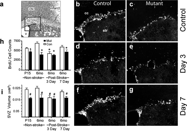
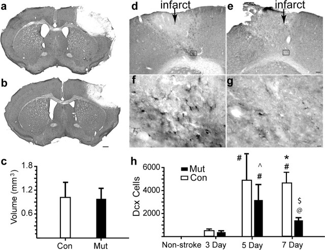
Erythropoietin (EPO) is the principal growth factor regulating the production of red blood cells. Recent studies demonstrated that exogenous EPO acts as a neuroprotectant and regulates neurogenesis. Using a genetic approach, we evaluate the roles of endogenous EPO and its classical receptor (EPOR) in mammalian neurogenesis. We demonstrate severe and identical embryonic neurogenesis defects in animals null for either the Epo or EpoR gene, suggesting that the classical EPOR is essential for EPO action during embryonic neurogenesis. Furthermore, by generating conditional EpoR knock-down animals, we demonstrate that brain-specific deletion of EpoR leads to significantly reduced cell proliferation in the subventricular zone and impaired post-stroke neurogenesis. EpoR conditional knockdown leads to a specific deficit in post-stroke neurogenesis through impaired migration of neuroblasts to the peri-infarct cortex. Our results suggest that both EPO and EPOR are essential for early embryonic neural development and that the classical EPOR is important for adult neurogenesis and for migration of regenerating neurons during post-injury recovery.


Figures

Figure 1.
Figure 1.
Epo and EpoR expression and neurogenesis defects in null animals. a–f, In situ hybridization of transverse sections with EpoR (a, c, e) or Epo (b, d, f) antisense probes on E8.5, E9.5, and E10.5 embryos. Inset photomicrographs are higher-magnification images from the neuroepithelium (a, c) and neural crest (e). g–l, Epo- (k, l) and EpoR- (g–j) deficient embryos show underdeveloped choroids plexus (i–l) and forebrain (g, h) at E13.5. Con, Control; Mut, mutant.
Figure 2.
Figure 2.
Analysis of EpoR conditional knock-down animals. a, PCR. Floxed, WT, and excised alleles (Δloxp) are indicated. b, RT-PCR analysis for EpoR expression levels. c, Hematocrit, brain weight, and body weight are unchanged in the mutants. Error bars indicate SDs. d, Cresyl violet sections from control (left) and mutant (right) animals. The bottom panels show higher-magnification views of the boxed areas. Con, Control; Mut, mutant.
Figure 3.
Figure 3.
Quantification of BrdU labeling in the SVZ. a, Cresyl violet section. The rectangle shows the location of b–g. b–g, BrdU-labeled cells in control (b, d, f) and EpoR conditional knock-down (c, e, g) animals in non-stroke (b, c), 3 d after stroke (d, e), and 7 d after stroke (f, g) are shown. Scale bar, 50 μm. h, i, The graphs show quantifications of BrdU-labeled cells (h) and SVZ volume before and after stroke (i). *p ≤ 0.004; #p ≤ 0.008; $p = 0.016. Error bars indicate SDs. ctx, Cortex; str, stiatum; V, ventricle; cc, corpus callosum; P15, postnatal day 15; mo, month; Con, control; Mut, mutant.
Figure 4.
Figure 4.
EpoR conditional knock-down leads to reduced neuroblast migration. a, b, Cresyl violet sections through the frontal cortex of mutant (a) and control (b) 7 d after stroke. Scale bar, 500 μm. c, Infarct volume 7 d after stroke was quantified. d–g, DCX+ cells in control (d, f) and mutant (e, g). The region within the box in d and e is enlarged in f and g. Scale bars: d, 100 μm; f, 25 μm. h, Stereological quantification of DCX+ cells. #p ≤ 0.001 versus all other conditions; *p = 0.008 versus Mut at non-stroke stage; ^p = 0.02 versus Mut at 7 d; @p < 0.02 versus 3 d Mut/Con and non-stroke; $p = 0.002 versus 7 d control. Error bars indicate SDs.

References

    1. Arvidsson A, Collin T, Kirik D, Kokaia Z, Lindvall O (2002). Neuronal replacement from endogenous precursors in the adult brain after stroke. Nat Med 8:963–970. - PubMed
    1. Bernaudin M, Marti HH, Roussel S, Divoux D, Nouvelot A, MacKenzie ET, Petit E (1999). A potential role for erythropoietin in focal permanent cerebral ischemia in mice. J Cereb Blood Flow Metab 19:643–651. - PubMed
    1. Brines M, Grasso G, Fiordaliso F, Sfacteria A, Ghezzi P, Fratelli M, Latini R, Xie QW, Smart J, Su-Rick CJ, Pobre E, Diaz D, Gomez D, Hand C, Coleman T, Cerami A (2004). Erythropoietin mediates tissue protection through an erythropoietin and common beta-subunit heteroreceptor. Proc Natl Acad Sci USA 101:14907–14912. - PMC - PubMed
    1. Brines ML, Ghezzi P, Keenan S, Agnello D, de Lanerolle NC, Cerami C, Itri LM, Cerami A (2000). Erythropoietin crosses the blood-brain barrier to protect against experimental brain injury. Proc Natl Acad Sci USA 97:10526–10531. - PMC - PubMed
    1. Carmichael ST (2005). Rodent stroke models: size, mechanism and purpose. NeuroRx 2:396–409. - PMC - PubMed

Publication types

MeSH terms

LinkOut - more resources