

Myeloid cells in infantile hemangioma
- PMID: 16436675
- PMCID: PMC1606494
- DOI: 10.2353/ajpath.2006.050618
Myeloid cells in infantile hemangioma
Abstract
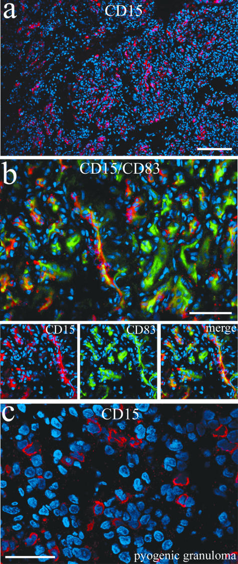
Little is known about the pathogenesis of infantile hemangiomas despite the fact that they are relatively common tumors. These benign neoplasms occur in as many as 1 in 10 births, and although rarely life threatening, hemangiomas can pose serious concerns to the cosmetic and psychosocial development of the afflicted child. Ulceration, scarring, and disfigurement are significant problems as are encroachment of the ear and eye, which can threaten hearing and vision. The precise mechanisms controlling the rapid growth observed in the first months of life and the spontaneous involution that follows throughout the course of years remain unknown. In this report we demonstrate the presence of large numbers of hematopoietic cells of the myeloid lineage in proliferating hemangiomas and propose a mechanism for the observed evolution of these lesions that is triggered by hypoxia and involves the participation of myeloid cells. We report the results of experiments using myeloid markers (CD83, CD32, CD14, CD15) that unexpectedly co-labeled hemangioma endothelial cells, providing new evidence that these cells are distinct from normal endothelium.
Figures


Similar articles
-
Identifying potential regulators of infantile hemangioma progression through large-scale expression analysis: a possible role for the immune system and indoleamine 2,3 dioxygenase (IDO) during involution.Lymphat Res Biol. 2003;1(4):291-9. doi: 10.1089/153968503322758094. Lymphat Res Biol. 2003. PMID: 15624557
-
Evaluation of expression profiles of hematopoietic stem cell, endothelial cell, and myeloid cell antigens in spontaneous and chemically induced hemangiosarcomas and hemangiomas in mice.Toxicol Pathol. 2013 Jul;41(5):709-21. doi: 10.1177/0192623312464309. Epub 2012 Nov 2. Toxicol Pathol. 2013. PMID: 23125116
-
The embryo-placental CD15-positive "vasculogenic zones" as a source of propranolol-sensitive pediatric vascular tumors.Placenta. 2016 Feb;38:93-9. doi: 10.1016/j.placenta.2015.12.013. Epub 2016 Jan 6. Placenta. 2016. PMID: 26907387
-
Evolution of hemangioma endothelium.Exp Mol Pathol. 2012 Oct;93(2):264-72. doi: 10.1016/j.yexmp.2012.04.020. Epub 2012 May 4. Exp Mol Pathol. 2012. PMID: 22565125 Review.
-
Signaling mechanisms in infantile hemangioma.Curr Opin Hematol. 2009 May;16(3):202-8. doi: 10.1097/MOH.0b013e32832a07ff. Curr Opin Hematol. 2009. PMID: 19367160 Free PMC article. Review.
Cited by
-
Beta-blockers as therapy for infantile hemangiomas.Semin Plast Surg. 2014 May;28(2):87-90. doi: 10.1055/s-0034-1376259. Semin Plast Surg. 2014. PMID: 25045334 Free PMC article.
-
Multipotential stem cells recapitulate human infantile hemangioma in immunodeficient mice.J Clin Invest. 2008 Jul;118(7):2592-9. doi: 10.1172/JCI33493. J Clin Invest. 2008. PMID: 18535669 Free PMC article.
-
Proliferating Infantile Hemangioma Tissues and Primary Cell Lines Express Markers Associated with Endothelial-to-Mesenchymal Transition.Plast Reconstr Surg Glob Open. 2020 Feb 11;8(2):e2598. doi: 10.1097/GOX.0000000000002598. eCollection 2020 Feb. Plast Reconstr Surg Glob Open. 2020. PMID: 32309069 Free PMC article.
-
Suppressed NFAT-dependent VEGFR1 expression and constitutive VEGFR2 signaling in infantile hemangioma.Nat Med. 2008 Nov;14(11):1236-46. doi: 10.1038/nm.1877. Epub 2008 Oct 19. Nat Med. 2008. PMID: 18931684 Free PMC article.
-
Expression of Cathepsins B, D, and G in Infantile Hemangioma.Front Surg. 2015 Jun 17;2:26. doi: 10.3389/fsurg.2015.00026. eCollection 2015. Front Surg. 2015. PMID: 26137466 Free PMC article.
References
-
- Mulliken JB, Glowacki J. Hemangiomas and vascular malformations in infants and children: a classification based on endothelial characteristics. Plast Reconstr Surg. 1982;69:412–422. - PubMed
-
- Tan ST, Wallis RA, He Y, Davis PF. Mast cells and hemangioma. Plast Reconstr Surg. 2004;113:999–1011. - PubMed
-
- Yu Y, Flint AF, Mulliken JB, Wu JK, Bischoff J. Endothelial progenitor cells in infantile hemangioma. Blood. 2004;103:1373–1375. - PubMed
-
- Dadras SS, North PE, Bertoncini J, Mihm MC, Detmar M. Infantile hemangiomas are arrested in an early developmental vascular differentiation state. Mod Pathol. 2004;17:1068–1079. - PubMed
Publication types
MeSH terms
Substances
Grants and funding
LinkOut - more resources
Full Text Sources
Medical
Research Materials
Miscellaneous
