. 2006 Feb;168(2):659-69.
doi: 10.2353/ajpath.2006.050599.

Hypoxia-induced pulmonary vascular remodeling requires recruitment of circulating mesenchymal precursors of a monocyte/macrophage lineage

Affiliations

Hypoxia-induced pulmonary vascular remodeling requires recruitment of circulating mesenchymal precursors of a monocyte/macrophage lineage

Maria G Frid et al. Am J Pathol. 2006 Feb.

Abstract

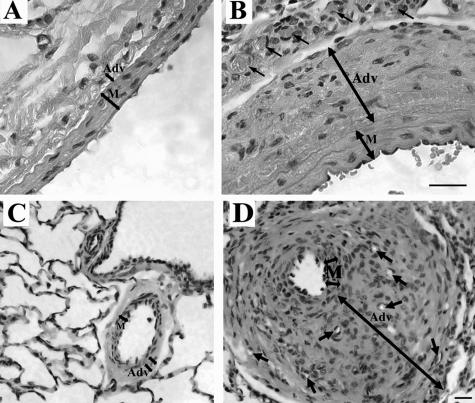
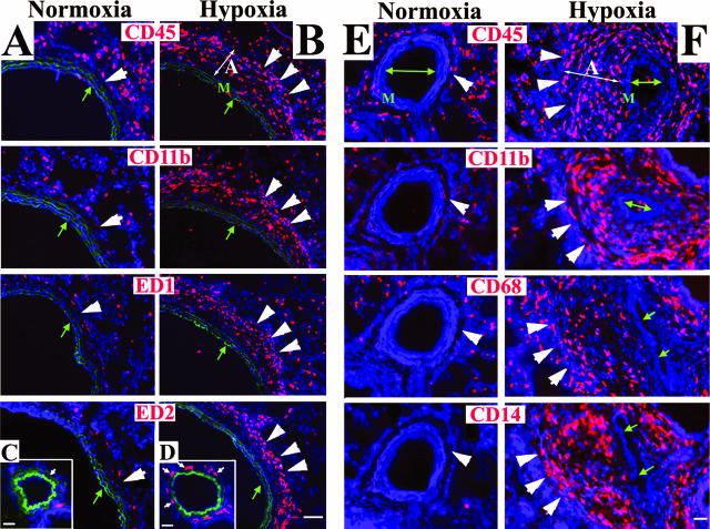
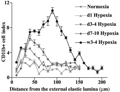
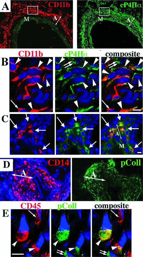
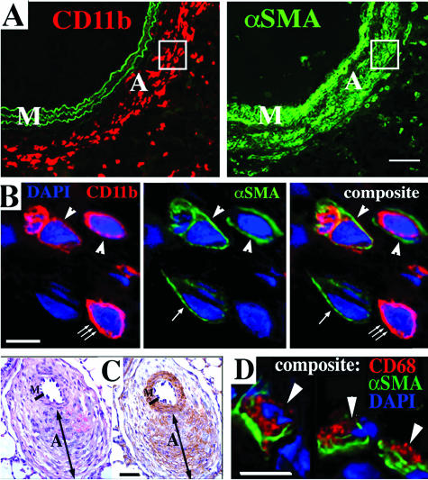
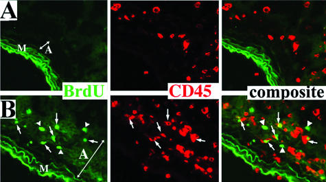
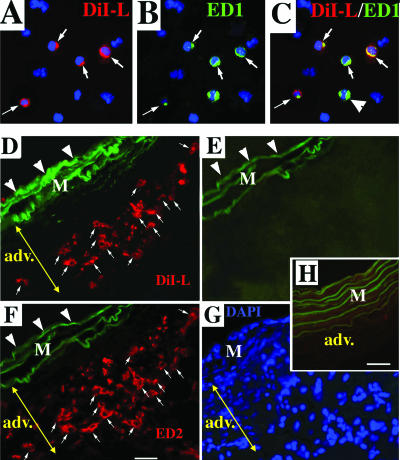
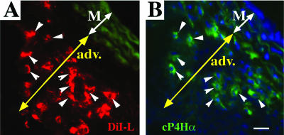
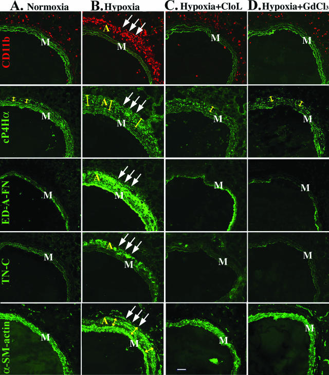
Vascular remodeling in chronic hypoxic pulmonary hypertension includes marked fibroproliferative changes in the pulmonary artery (PA) adventitia. Although resident PA fibroblasts have long been considered the primary contributors to these processes, we tested the hypothesis that hypoxia-induced pulmonary vascular remodeling requires recruitment of circulating mesenchymal precursors of a monocyte/macrophage lineage, termed fibrocytes. Using two neonatal animal models (rats and calves) of chronic hypoxic pulmonary hypertension, we demonstrated a dramatic perivascular accumulation of mononuclear cells of a monocyte/macrophage lineage (expressing CD45, CD11b, CD14, CD68, ED1, ED2). Many of these cells produced type I collagen, expressed alpha-smooth muscle actin, and proliferated, thus exhibiting mesenchymal cell characteristics attributed to fibrocytes. The blood-borne origin of these cells was confirmed in experiments wherein circulating monocytes/macrophages of chronically hypoxic rats were in vivo-labeled with DiI fluorochrome via liposome delivery and subsequently identified in the remodeled pulmonary, but not systemic, arterial adventitia. The DiI-labeled cells that appeared in the vessel wall expressed monocyte/macrophage markers and procollagen. Selective depletion of this monocytic cell population, using either clodronate-liposomes or gadolinium chloride, prevented pulmonary adventitial remodeling (ie, production of collagen, fibronectin, and tenascin-C and accumulation of myofibroblasts). We conclude that circulating mesenchymal precursors of a monocyte/macrophage lineage, including fibrocytes, are essential contributors to hypoxia-induced pulmonary vascular remodeling.


Figures

Figure 1
Figure 1
Chronic hypoxia induces PA adventitial thickening. H&E staining of pulmonary arteries from rats (A, B) and calves (C, D). The adventitial layer (Adv) is very thin in normoxic rats (A) and calves (C). In chronically hypoxic animals of both species, the PA adventitia is remarkably thickened (B, rats; D, calves). Small arrows point to newly formed capillaries of the vasa vasorum. M, tunica media. Scale bars, 20 μm.
Figure 2
Figure 2
Hypoxia induces a robust appearance of MNCs in the PA adventitia. Cryosections of PAs from rats (A–D) and calves (E, F) were labeled with antibodies against MNC/macrophage antigens (red) and cell nuclei (DAPI, blue). Few MNCs are present in the adventitia of normoxic rats and calves (single large arrowheads in the normoxia columns). In the adventitia of chronically hypoxic rats and calves, abundant MNCs of a monocyte/macrophage phenotype are present (triple large arrowheads in the hypoxia columns). For rats, micrographs A and B show main PAs, whereas C and D show distal PAs (80 to 100 μm in diameter). Green autofluorescence of the elastic lamellae in A--D defines the boundaries of the PA tunica media. The PA lumens are marked with small green arrows. M, media; A, adventitia. Scale bars: 100 μm (A, B); 20 μm (C–F).
Figure 3
Figure 3
Chronic hypoxia causes a progressive accumulation of MNCs in the PA adventitia. Time course analysis of accumulation of MNCs expressing CD11b antigen (CD11b+ cell index) in the PA adventitia of chronically hypoxic (1 day to 4 weeks) rats demonstrates a significant and progressive increase in the CD11b+ cell index throughout time, compared to normoxic controls. Furthermore, a progressive increase in the distance from the external elastic lamellae (x axis) of the CD11b+ cell peak was observed that correlated with the progressive increase in the thickening of PA adventitia in response to chronic exposure to hypoxia.
Figure 4
Figure 4
Monocytic cells in the remodeled PA adventitia express procollagen. A: Serial tissue sections of the rat PA labeled with antibodies against a monocytic cell marker CD11b (red, left) and collagen precursor molecule, collagen-prolyl-hydroxylase-α (cP4Hα) (green, right). Elastic lamellae of the tunica media (left) exhibit green autofluorescence, which is shown to better define the tunica media boundaries. B: Micrograph of the adventitial area shown within the frame in A. Double-label immunofluorescent staining followed by deconvolution confocal microscopy demonstrates that the remodeled PA adventitia of chronically hypoxic rats contains numerous cells that co-express (arrowheads) CD11b (red fluorescence) and cP4Hα (green fluorescence). There is a cell (small single arrow) that expresses a monocytic antigen CD11b (red) but not cP4Hα, and another cell (small double arrow) expresses cP4Hα but not CD11b. DAPI (blue) labels all cell nuclei. C: Routine immunofluorescence microscopy of the small distal PA of chronically hypoxic rat shows adventitial cells that also co-express CD11b (red) and cP4Hα (green) (arrows). D: Serial sections of the distal PA of chronically (2 week) hypoxic calf was labeled with CD14 Abs (red) and type I procollagen (pColl) Abs (green). E: Double-label immunofluorescent staining followed by deconvolution confocal microscopy demonstrates adventitial cells from the 2-week hypoxic calf that co-express (arrowheads) CD45 (red) and pColl (green). There is also a cell that expresses CD45 but not pColl (small single arrow), and a cell that expresses pColl but not CD45 (small double arrows). M, media; A, adventitia. Scale bars, 5 μm.
Figure 5
Figure 5
Monocytic cells in the remodeled PA adventitia express α-SMA. A: Serial tissue sections of the rat PA labeled with antibodies against a monocytic cell marker CD11b (red, left) and α-SMA (αSMA, green, right). Elastic lamellae of the tunica media (left) exhibit green autofluorescence, which is shown to better define the tunica media boundaries. B: Micrograph of the adventitial area shown within the frame in A. Double-label immunofluorescent staining and deconvolution confocal microscopy demonstrate adventitial cells that co-express (arrowheads) CD11b (red) and α-SMA (αSMA, green). There is a cell (small triple arrow) that expresses a monocytic antigen CD11b (red) but not α-SMA, and another cell (small single arrow) expresses α-SMA but not CD11b. DAPI (blue) labels cell nuclei. C: Distal PA of the chronically (2 week) hypoxic calf was labeled with H&E (left) or with peroxidase-conjugated antibodies against α-SMA (brown, right). PA adventitia is remarkably thickened (similar to image in Figure 1D), and α-SMA immunoreactivity (brown) is observed not only in the tunica media but also in some areas of the adventitia. D: Double-label immunofluorescent staining and deconvolution confocal microscopy demonstrate cells from the adventitia of a hypoxic calf that co-express (arrowheads) a macrophage marker CD68 (red) and α-SMA (green). DAPI (blue) labels cell nuclei. M, media; A, adventitia. Scale bars: 50 μm (A, C); 5 μm (B, D).
Figure 6
Figure 6
Monocytic cells actively replicate in the remodeled PA adventitia. Double-label immunofluorescent staining of the remodeled PA from the chronically (72 hours) hypoxic rats (B) demonstrates numerous adventitial cells that co-express (arrows) MNC antigens (CD45, red) and proliferation-associated marker, BrdU (green). Several cells that have also incorporated BrdU, but do not express MNC antigen, CD45, are marked by arrowheads. Adventitial cells in the PA of age-matched normoxic rats (A) do not demonstrate BrdU incorporation. Elastic lamellae of the PA media exhibit green autofluorescence and are shown to define the medial-adventitial border. M, media; A, adventitia.
Figure 7
Figure 7
Fibrocytes, which have accumulated in the remodeled PA adventitia, originate from the circulation. A–C: Cytospins of MNCs isolated from peripheral blood of rats that were exposed to hypoxia for 96 hours and were intravenously injected with DiI liposomes 2 hours before drawing blood. A: MNCs that phagocytized DiI liposomes (DiI-L) exhibit red fluorescence. B: MNCs labeled with antibodies against a monocytic marker ED1 exhibit green fluorescence. C: Composite image demonstrates that DiI liposomes are taken up in the circulation by MNCs of exclusively monocytic lineage (ED1+) (small arrows). An arrowhead points to one of the ED1+ MNCs that was not DiI-labeled in the circulation. D: Monocytic cells labeled in the circulation with DiI (red fluorochrome) via liposome-mediated delivery (see Materials and Methods and A–C above) are subsequently observed in the remodeled PA adventitia of chronically (4 week) hypoxic rats (small arrows). E and F: The DiI+ cells (D) express monocyte/macrophage phenotype. E and F: After the images of DiI+ cells (seen in D) were acquired, DiI fluorescence was completely bleached by fixing this tissue cryosection in acetone/methanol (E), and next immunolabeling for a macrophage marker ED2 (red) was performed on the same cryosection (F). Small arrows in D and F point to numerous cells that show both DiI-labeling (D) and ED2 expression (F). Large arrowheads in D–F point to elastic lamellae of the tunica media (M), which exhibit green autofluorescence (shown to define the boundaries of the medial layer). Yellow double-headed arrows mark the PA adventitia (adv). G: Cell nuclei on the same tissue section are shown by DAPI immunoreactivity (blue). H: Aorta from the chronically hypoxic rat (same one as in D–G) that was intravenously injected with DiI liposomes, demonstrates the absence of DiI+ cells. Scale bars, 10 μm.
Figure 8
Figure 8
Circulating MNCs that have accumulated in the remodeled PA adventitia express a fibrocyte phenotype. A and B: MNCs that were labeled with DiI (red fluorescence) in the circulation (see Materials and Methods and Figure 7, A–C), are subsequently found in the remodeled PA adventitia of chronically (4 week) hypoxic rat (A), and express cP4Hα (procollagen) (green fluorescence) (B). Arrowheads point to fibrocytes that demonstrate both DiI-labeling and expression of cP4Hα. M, media; A, adventitia. Scale bars, 20 μm.
Figure 9
Figure 9
Depletion of fibrocytes in the circulation of chronically hypoxic rats attenuates PA adventitial remodeling. A: Normoxia column: row CD11b: the PA adventitia of normoxic rats demonstrates few CD11b+ MNCs (red). Elastic lamellae of the tunica media exhibit green autofluorescence and are shown to define the boundaries of the tunica media. Row cP4Hα: The PA adventitia of normoxic rats is comprised of resident adventitial fibroblasts expressing cP4Hα (green fluorescence within the adventitia). Adventitia in normoxic rats is thin (yellow arrows). Rows ED-A-FN and TN-C: Resident fibroblasts in the PA adventitia of normoxic rats do not express cellular ED-A-fibronectin (ED-A-FN) or tenascin-C (TN-C). Row α-SM-actin: Resident fibroblasts in the PA adventitia of normoxic rats do not express α-SMA [SMC of the tunica media (M) express α-SMA]. B: Hypoxia column: rows CD11b and cP4Hα: the remodeled PA adventitia (triple arrows) of chronically (4 week) hypoxic rats demonstrates numerous CD11b+ cells (row CD11b, red), and markedly thickened adventitia (marked by yellow arrows in row cP4Hα) with numerous cP4Hα-expressing cells (green fluorescence within the adventitia). Rows ED-A-FN and TN-C: The remodeled PA adventitia of chronically hypoxic rats demonstrates deposition of ED-A-fibronectin and tenascin-C (rows ED-A-FN and TN-C, green fluorescence). Row α-SM-actin: Numerous α-SMA-expressing myofibroblasts are observed in the PA adventitia of chronically hypoxic rats (triple arrows). C and D: Hyp+Cl2MBP-L and Hyp+GdCl3 columns: selective depletion (see Materials and Methods and Results sections) of monocytic cells in the circulation of chronically hypoxic rats by either clodronate-liposomes (C, Hypoxia+Cl2MBP-L) or gadolinium chloride (D, Hypoxia+GdCl3) results in diminished accumulation of CD11+ cells in the PA adventitia (row CD11b, red), reduced adventitial thickness (row cP4Hα, yellow arrows), prevention of adventitial fibrosis [absence of fibronectin (row ED-A-FN) and tenascin-C (row TN-C) deposition], as well as prevention of α-SMA expression by adventitial fibroblasts (row α-SMA). M, tunica media. Scale bar, 100 μm.

References

    1. Stenmark KR, Mecham R. Cellular and molecular mechanisms of pulmonary vascular remodeling. Annu Rev Physiol. 1997;59:89–144. - PubMed
    1. Jeffery T, Morrell NW. Molecular and cellular basis of pulmonary vascular remodeling in pulmonary hypertension. Prog Cardiovasc Dis. 2002;45:173–202. - PubMed
    1. Rabinovitch M. Pulmonary vascular remodeling in hypoxic pulmonary hypertension. Yuan JX-J, editor. Dordrecht: Kluwer Academic Publishers,; Hypoxic Pulmonary VasoconstrictionCellular and Molecular Mechanisms. 2004:pp 403–418.
    1. Meyrick B. The pathology of pulmonary artery hypertension. Clin Chest Med. 2001;22:393–404. - PubMed
    1. Humbert M, Morrell NW, Archer SL, Stenmark KR, MacLean MR, Lang IM, Christman BW, Weir EK, Eickelberg O, Voelkel NF, Rabinovitch M. Cellular and molecular pathobiology of pulmonary arterial hypertension. J Am Coll Cardiol. 2004;43(Suppl S):13S–24S. - PubMed

Publication types

MeSH terms