. 2006 Feb;168(2):695-705.
doi: 10.2353/ajpath.2006.050759.

Vascular endothelial growth factor antagonist modulates leukocyte trafficking and protects mouse livers against ischemia/reperfusion injury

Affiliations

Vascular endothelial growth factor antagonist modulates leukocyte trafficking and protects mouse livers against ischemia/reperfusion injury

Sei-ichiro Tsuchihashi et al. Am J Pathol. 2006 Feb.

Abstract

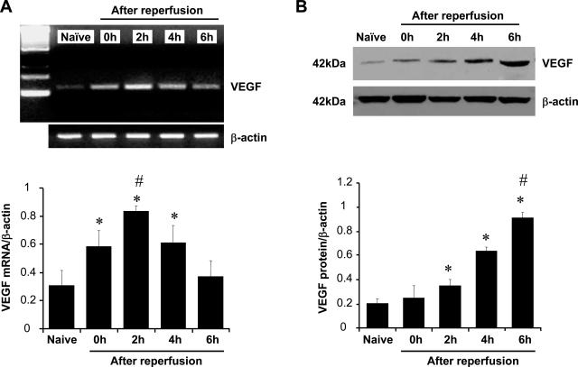
Although hypoxia stimulates the expression of vascular endothelial growth factor (VEGF), little is known of the role or mechanism by which VEGF functions after ischemia and reperfusion (I/R) injury. In this report, we first evaluated the expression of VEGF in a mouse model of liver warm ischemia. We found that the expression of VEGF increased after ischemia but peaked between 2 and 6 hours after reperfusion. Mice were treated with a neutralizing anti-mouse VEGF antiserum (anti-VEGF) or control serum daily from day -1 (1 day before the initiation of ischemia). Treatment with anti-VEGF significantly reduced serum glutaminic pyruvic transaminase levels and reduced histological evidence of hepatocellular damage compared with controls. Anti-VEGF also markedly decreased T-cell, macrophage, and neutrophil accumulation within livers and reduced the frequency of intrahepatic apoptotic terminal deoxynucleotidyl transferase-mediated dUTP nick end-labeling-positive cells. Moreover, there was a reduction in the expression of pro-inflammatory cytokines (tumor necrosis factor-alpha and interferon-gamma), chemokines (interferon-inducible protein-10 and monocyte chemoattractant protein-1) and adhesion molecules (E-selectin) in parallel with enhanced expression of anti-apoptotic genes (Bcl-2/Bcl-xl and heme oxygenase-1) in anti-VEGF-treated animals. In conclusion, hypoxia-inducible VEGF expression by hepatocytes modulates leukocyte trafficking and leukocyte-induced injury in a mouse liver model of warm I/R injury, demonstrating the importance of endogenous VEGF production in the pathophysiology of hepatic I/R injury.


Figures

Figure 1
Figure 1
Generation of anti-VEGF antiserum. A: Every 6 weeks, after immunization with VEGF peptide, New Zealand White rabbits were bled, and serum was collected. An ELISA is illustrated for the assessment of anti-VEGF in four postimmunization immune sera compared with pre-immune serum. ELISA was performed using 2 μg/ml VEGF peptide as the antigen and a goat anti-rabbit horseradish peroxidase-conjugated antibody as the secondary reagent. B: Human endothelial cells were treated with mouse VEGF (5 ng/ml) in triplicate cultures, and after 72 hours, proliferation was assessed by 3[H]thymidine incorporation. Anti-VEGF antiserum was added to cultures at increasing dilutions, as illustrated. The percent inhibition of mean VEGF-induced proliferation was calculated for each culture condition. No inhibition was found with control serum at identical concentrations (not shown). One representative experiment of 10 is shown.
Figure 2
Figure 2
Kinetics of VEGF expression in mouse liver after 90 minutes of warm ischemia followed by 6 hours of reperfusion. A: Competitive-template RT-PCR analysis. The expression of VEGF mRNA was significantly up-regulated after 90 minutes of ischemia (0 hours) and peaked at 2 hours of reperfusion compared with naïve control liver. It decreased at 6 hours after reperfusion (mean ± SD; n = 4/time-point). *P < 0.05 versus naïve, #P < 0.05 versus 0 or 4 hours. B: Western blot analysis. The expression of VEGF protein was significantly up-regulated beginning at 2 hours of reperfusion with peak up-regulation taking place at 6 hours of reperfusion (mean ± SD; n = 4). *P < 0.01 versus naïve, #P < 0.01 versus 0, 2, and 4 hours.
Figure 3
Figure 3
Serum GPT levels (IU/L). The sGPT levels were significantly lower in anti-VEGF serum-treated mice compared with control mice (mean ± SD; n = 4–9/group). *P < 0.01 versus control group.
Figure 4
Figure 4
Photomicrograms of representative mouse livers. A and B: Sham group. C and D: Control group with severe sinusoidal congestion and hepatocyte necrosis (Suzuki’s score = 7.97 ± 1.08). E and F: Anti-VEGF group with good preservation of lobular architecture without edema, congestion, or absence of centrilobular necrosis (Suzuki’s score = 1.00 ± 0.87). H&E stain; representative of four to nine per group.
Figure 5
Figure 5
VEGF mRNA/protein expression after anti-VEGF serum treatment (at 6 hours of reperfusion). A: Competitive-template RT-PCR analysis. No significant differences in mRNA coding for VEGF were observed among sham, control, and treatment groups (mean ± SD; n = 4–6/group). B: Western blot analysis. The expression of VEGF protein was significantly increased in the control and anti-VEGF serum treatment groups compared with sham group. Anti-VEGF decreased VEGF protein synthesis compared with controls. Results are representative of four different experiments. *P < 0.01 versus sham. #P < 0.01.
Figure 6
Figure 6
Intrahepatic neutrophil accumulation. MPO enzyme activity levels (U/g) were significantly lower in anti-VEGF serum treatment group compared with control group. These data represent the mean ± SD of four to six experiments. *P < 0.05.
Figure 7
Figure 7
Immunohistochemical staining for intrahepatic leukocytes (CD45), T cells (CD3), and macrophages (Mac). Control serum-treated mice with increased intrahepatic leukocyte (B), T-cell (E), and macrophage (H) infiltration. In contrast, mice treated with anti-VEGF Ab revealed minimal leukocyte (C), T-cell (F), and macrophage (I) infiltration. Representative of four animals per group. Original magnification, ×400.
Figure 8
Figure 8
Competitive template RT-PCR-assisted expression of mRNA coding for TNF-α, IFN-γ, IP-10, MCP-1, and E-selectin. The control group shows significant up-regulation of pro-inflammatory cytokines (TNF-α and IFN-γ), chemokines (IP-10 and MCP-1), and E-selectin expression compared with those treated with anti-VEGF Ab. These data represent the mean ± SD of four to six experiments. *P < 0.05.
Figure 9
Figure 9
TUNEL-assisted detection of apoptosis. A: Sham group. B: Control group with large numbers of TUNEL+ cells (dark brown spots). C: Anti-VEGF group with markedly decreased frequency of TUNEL+ cells. D: The results were scored semiquantitatively by averaging the number of TUNEL+ cells (mean ± SD) per microscopic field at magnification ×400. Minimum of 10 fields were evaluated per sample in four different experiments. *P < 0.01.
Figure 10
Figure 10
Western blot-assisted analysis of anti-apoptotic (Bcl-2/Bcl-xl), pro-apoptotic (Bax), and antioxidant (HO-1) proteins. A: Bcl-2, Bcl-xl, Bax, and HO-1 proteins were detected by polyclonal rabbit anti-mouse Abs. Antibody against β-actin was used as internal control. B: Each bar graph shows the ratio of protein and β-actin expression. Bcl-2/Bcl-xl and HO-1 expression was increased in anti-VEGF group compared with controls. The expression of Bax was suppressed by VEGF antagonist. Results are representative of four different experiments. *P < 0.01.

References

    1. Farmer DG, Amersi F, Busuttil RW, Kupiec-Weglinski JW. Current concepts in ischemia and reperfusion injury in the liver. Transplant Rev. 2000;14:106–126.
    1. Fondevila C, Busuttil RW, Kupiec-Weglinski JW. Hepatic ischemia/reperfusion injury: a fresh look. Exp Mol Pathol. 2003;74:86–93. - PubMed
    1. Teoh NC, Farrell GC. Hepatic ischemia reperfusion injury: pathogenic mechanisms and basis for hepatoprotection. J Gastroenterol Hepatol. 2003;18:891–902. - PubMed
    1. Reinders ME, Sho M, Izawa A, Wang P, Mukhopadhyay D, Koss KE, Geehan CS, Luster AD, Sayegh MH, Briscoe DM. Proinflammatory functions of vascular endothelial growth factor in alloimmunity. J Clin Invest. 2003;112:1655–1665. - PMC - PubMed
    1. Levy AP, Levy NS, Wegner S, Goldberg MA. Transcriptional regulation of the rat vascular endothelial growth factor gene by hypoxia. J Biol Chem. 1995;270:13333–13340. - PubMed

Publication types

MeSH terms