. 2006 Feb;50(2):639-48.
doi: 10.1128/AAC.50.2.639-648.2006.

Potencies of human immunodeficiency virus protease inhibitors in vitro against Plasmodium falciparum and in vivo against murine malaria

Affiliations

Potencies of human immunodeficiency virus protease inhibitors in vitro against Plasmodium falciparum and in vivo against murine malaria

Katherine T Andrews et al. Antimicrob Agents Chemother. 2006 Feb.

Abstract

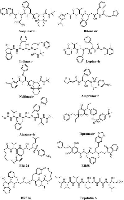
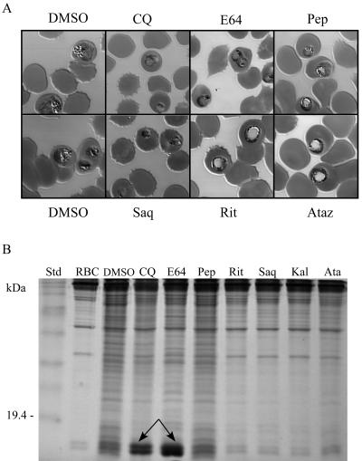
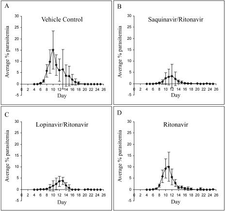
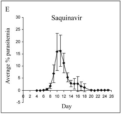
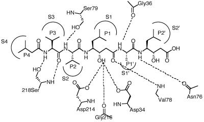
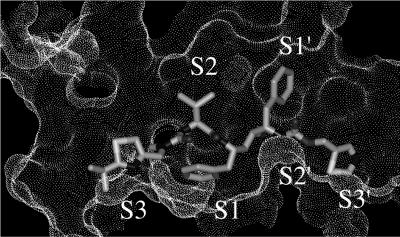
Parasite resistance to antimalarial drugs is a serious threat to human health, and novel agents that act on enzymes essential for parasite metabolism, such as proteases, are attractive targets for drug development. Recent studies have shown that clinically utilized human immunodeficiency virus (HIV) protease inhibitors can inhibit the in vitro growth of Plasmodium falciparum at or below concentrations found in human plasma after oral drug administration. The most potent in vitro antimalarial effects have been obtained for parasites treated with saquinavir, ritonavir, or lopinavir, findings confirmed in this study for a genetically distinct P. falciparum line (3D7). To investigate the potential in vivo activity of antiretroviral protease inhibitors (ARPIs) against malaria, we examined the effect of ARPI combinations in a murine model of malaria. In mice infected with Plasmodium chabaudi AS and treated orally with ritonavir-saquinavir or ritonavir-lopinavir, a delay in patency and a significant attenuation of parasitemia were observed. Using modeling and ligand docking studies we examined putative ligand binding sites of ARPIs in aspartyl proteases of P. falciparum (plasmepsins II and IV) and P. chabaudi (plasmepsin) and found that these in silico analyses support the antimalarial activity hypothesized to be mediated through inhibition of these enzymes. In addition, in vitro enzyme assays demonstrated that P. falciparum plasmepsins II and IV are both inhibited by the ARPIs saquinavir, ritonavir, and lopinavir. The combined results suggest that ARPIs have useful antimalarial activity that may be especially relevant in geographical regions where HIV and P. falciparum infections are both endemic.


Figures

FIG. 1.
FIG. 1.
Structures of aspartyl protease inhibitors.
FIG. 2.
FIG. 2.
Effect of protease inhibitors on P. falciparum development and hemoglobin digestion. Ring-stage in vitro-cultured Dd2-parasitized erythrocytes were incubated for 20 h with either 1 μM chloroquine or 10 μM E64, pepstatin A (Pep), ritonavir (Rit), saquinavir (Saq), atazanavir (Ataz), or ritonavir-lopinavir (Kaletra [Kal]). In parallel controls, parasites were treated with an equivalent concentration of DMSO drug solvent. (A) Micrographs of representative Giemsa-stained parasitized erythrocytes. (B) Effect of protease inhibitors on P. falciparum hemoglobin hydrolysis (15% sodium dodecyl sulfate-polyacrylamide gel electrophoresis). Arrows indicate inhibition of hemoglobin digestion by chloroquine (CQ; partial) and E64. RBC, uninfected red blood cells.
FIG. 3.
FIG. 3.
In vivo efficacy of HIV-1 protease inhibitors against murine malaria. Female C57BL/6J mice infected with 105 P. chabaudi AS-parasitized erythrocytes were treated orally, twice daily, for 8 consecutive days starting 24 h p.i. with either (A) vehicle, (B) 10 mg/kg each ritonavir and saquinavir, (C) 10 mg/kg ritonavir and 40 mg/kg lopinavir, (D) 10 mg/kg ritonavir, or (E) 10 mg/kg saquinavir. Parasitemia was monitored for 25 days from day 4 p.i. No parasitemia developed in Malarone-treated mice (not shown).
FIG. 3.
FIG. 3.
In vivo efficacy of HIV-1 protease inhibitors against murine malaria. Female C57BL/6J mice infected with 105 P. chabaudi AS-parasitized erythrocytes were treated orally, twice daily, for 8 consecutive days starting 24 h p.i. with either (A) vehicle, (B) 10 mg/kg each ritonavir and saquinavir, (C) 10 mg/kg ritonavir and 40 mg/kg lopinavir, (D) 10 mg/kg ritonavir, or (E) 10 mg/kg saquinavir. Parasitemia was monitored for 25 days from day 4 p.i. No parasitemia developed in Malarone-treated mice (not shown).
FIG. 4.
FIG. 4.
Comparison of active sites of P. falciparum plasmepsin II (PDB, 1LF3) and P. chabaudi plasmepsin (homology model) showing side chain variations. Pepstatin A residues (P) are shown bound to P. falciparum plasmepsin II binding sites (S). Prime and nonprime designations distinguish C-terminal from N-terminal sides of the cleavage site. Subsite pocket residue differences in P. falciparum versus P. chabaudi (residues in italics) are as follows: S4 (I290T), S3 (T114I), S2 (L292V), S1 (F111L), S1′ (V78G; N76V), S2′ (N76V) (all residues numbered according to PfPM-II numbering).
FIG. 5.
FIG. 5.
Comparison of superimposed crystal structures of ligand-protease complexes (proteases not shown) for HIV-1 protease and P. falciparum plasmepsin II. (Top) Superimposition of crystal structures for HIV protease inhibitor complexes showing protease-bound ligand conformations: lopinavir (PDB code, 1mui), indinavir (1hsg), ritonavir (1hxw), amprenavir (1hpv), saquinavir (1hxb), nelfinavir (1ohr), BR124 (1d4l), BR314 (1d4k). Overlay was performed using backbone atoms N, Cα, and C (or equivalent) from P2-P1′ using the Search and Compare module within InsightII. (Middle) Superimposition of crystal structures for P. falciparum PM-II inhibitor complexes showing protease-bound conformations: pepstatin A (1m43), pepstatin A (1sme), statine-based ligand (1me6), rs367 (1lee), rs370 (1lf2), EH58 (1lf3). Overlay was performed using backbone atoms N, Cα, and C (or equivalent) from P3-P1′. (Bottom) Superimposition of above enzyme-bound HIV-1 protease inhibitors (black) on PfPM-II inhibitors (gray) using backbone atoms N, Cα, and C (or equivalent) from P2-P1′.
FIG. 6.
FIG. 6.
Connolly surface of active site of PfPM-II (1lf3) in complex with docked ritonavir showing enzyme subsites S3-S2′.

References

    1. Abbenante, G., and D. P. Fairlie. 2005. Protease inhibitors in the clinic. Med. Chem. 1:71-104. - PubMed
    1. Andrews, K. T., A. Walduck, M. J. Kelso, D. P. Fairlie, A. Saul, and P. G. Parsons. 2000. Anti-malarial effect of histone deacetylation inhibitors and mammalian tumour cytodifferentiating agents. Int. J. Parasitol. 30:761-768. - PubMed
    1. Asojo, O. A., S. V. Gulnik, E. Afonina, B. Yu, J. A. Ellman, T. S. Haque, and A. M. Silva. 2003. Novel uncomplexed and complexed structures of plasmepsin II, an aspartic protease from Plasmodium falciparum. J. Mol. Biol. 327:173-181. - PubMed
    1. Back, D. J., S. H. Khoo, S. E. Gibbons, and C. Merry. 2001. The role of therapeutic drug monitoring in treatment of HIV infection. Br. J. Clin. Pharmacol. 52:89S-96S. - PMC - PubMed
    1. Bailey, E., R. Jambou, J. Savel, and G. Jaureguiberry. 1992. Plasmodium falciparum: differential sensitivity in vitro to E-64 (cysteine protease inhibitor) and pepstatin A (aspartyl protease inhibitor). J. Protozool. 39:593-599. - PubMed

Publication types