Review
. 2006 Jan 25;34(2):635-46.
doi: 10.1093/nar/gkj469. Print 2006.

Experimental approaches to identify non-coding RNAs

Affiliations
Review

Experimental approaches to identify non-coding RNAs

Alexander Hüttenhofer et al. Nucleic Acids Res. .

Abstract

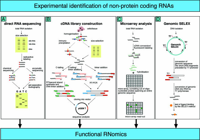
Cellular RNAs that do not function as messenger RNAs (mRNAs), transfer RNAs (tRNAs) or ribosomal RNAs (rRNAs) comprise a diverse class of molecules that are commonly referred to as non-protein-coding RNAs (ncRNAs). These molecules have been known for quite a while, but their importance was not fully appreciated until recent genome-wide searches discovered thousands of these molecules and their genes in a variety of model organisms. Some of these screens were based on biocomputational prediction of ncRNA candidates within entire genomes of model organisms. Alternatively, direct biochemical isolation of expressed ncRNAs from cells, tissues or entire organisms has been shown to be a powerful approach to identify ncRNAs both at the level of individual molecules and at a global scale. In this review, we will survey several such wet-lab strategies, i.e. direct sequencing of ncRNAs, shotgun cloning of small-sized ncRNAs (cDNA libraries), microarray analysis and genomic SELEX to identify novel ncRNAs, and discuss the advantages and limits of these approaches.


Figures

Figure 1
Figure 1
Four experimental approaches (A–D) to identify candidates for ncRNAs are shown. (A) Identification of ncRNAs by chemical or enzymatic sequencing of extracted abundant RNAs. (B) Identification of ncRNAs by cDNA cloning and sequencing; three different methods are indicated to reverse transcribe ncRNAs, usually lacking poly(A) tails, into cDNAs (e.g. by C-tailing, C-tailing and linker addition, or linker addition, only, followed by RT–RCR). (C) Identification of ncRNAs by micro-array analysis. DNA oligonucleotide covering the sequence space of an entire genome are spotted onto glass slides, to which fluorescently labelled samples, derived from cellular RNA, is hybridized. (D) Identification of ncRNAs by genomic SELEX. By random priming, the sequence of a genome is converted into short PCR fragments containing a T7 promotor at their 5′ ends. Subsequently, in vitro transcription by means of T7 RNA polymerase converts this genomic sequence of an organism into RNA fragments, which can then be assayed for function, such as binding to a specific protein or small chemical ligand, by SELEX.
Figure 2
Figure 2
Microarray detection of cellular RNAs, including ncRNAs, that associate with the bacterial Sm-like protein, Hfq, and the La homologous protein (Lhp1p) of yeast (S.cerevisiae), respectively (77,87). Left panel: RNAs are co-immunoprecipitated from E.coli cell extracts with anti-Hfq antibodies, purified and hybridized to high-density microarrays that carry DNA oligonucleotides covering the entire E.coli genome. RNAs that hybridized to probes on the arrays are detected with an antibody that specifically sees DNA:RNA hybrids. Subsequently, such signals are detected as indicated and subtracted from those obtained in a control experiment in which cell extracts were incubated with pre-immune sera, that is no immunoprecipitation of Hfq. Right panel: cell extracts from either wild-type yeast or yeast cells that express epitope (myc)-tagged Lhp1p are incubated with an anti-myc antibody, RNA is extracted from immunoprecipitates and reverse transcribed. The two obtained cDNAs are labelled with different fluorescent dyes (Cy3, red; Cy5, green), mixed and hybridized to yeast whole-genome microarrays. Spots that yield red signals indicate that the corresponding RNA was enriched in Lhp1p-myc immunoprecipitates.

References

    1. Mattick J.S. Non-coding RNAs: the architects of eukaryotic complexity. EMBO Rep. 2001;2:986–991. - PMC - PubMed
    1. Eddy S.R. Non-coding RNA genes and the modern RNA world. Nature Rev. Genet. 2001;2:919–929. - PubMed
    1. Huttenhofer A., Brosius J., Bachellerie J.P. RNomics: identification and function of small, non-messenger RNAs. Curr. Opin. Chem. Biol. 2002;6:835–843. - PubMed
    1. Huttenhofer A., Schattner P., Polacek N. Non-coding RNAs: hope or hype? Trends Genet. 2005;21:289–297. - PubMed
    1. Storz G. An expanding universe of noncoding RNAs. Science. 2002;296:1260–1263. - PubMed

Publication types

MeSH terms

Substances