. 2006 Feb 8;25(3):479-89.
doi: 10.1038/sj.emboj.7600948. Epub 2006 Jan 26.

Growth factor pleiotropy is controlled by a receptor Tyr/Ser motif that acts as a binary switch

Affiliations

Growth factor pleiotropy is controlled by a receptor Tyr/Ser motif that acts as a binary switch

Mark A Guthridge et al. EMBO J. .

Abstract

Pleiotropism is a hallmark of cytokines and growth factors; yet, the underlying mechanisms are not clearly understood. We have identified a motif in the granulocyte macrophage-colony-stimulating factor receptor composed of a tyrosine and a serine residue that functions as a binary switch for the independent regulation of multiple biological activities. Signalling occurs either through Ser585 at lower cytokine concentrations, leading to cell survival only, or through Tyr577 at higher cytokine concentrations, leading to cell survival as well as proliferation, differentiation or functional activation. The phosphorylation of Ser585 and Tyr577 is mutually exclusive and occurs via a unidirectional mechanism that involves protein kinase A and tyrosine kinases, respectively, and is deregulated in at least some leukemias. We have identified similar Tyr/Ser motifs in other cell surface receptors, suggesting that such signalling switches may play important roles in generating specificity and pleiotropy in other biological systems.


Figures
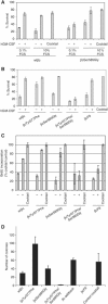
Figure 1
Figure 1
Tyr577 and Ser585 constitute a distinct motif that is essential for the regulation of cell survival and proliferation. Fetal liver cells from βc−/− βIL-3−/− double-knockout mice were transduced with wtβc and mutant receptor constructs. Following transduction, cells were plated in either no factor (−), 3.3 nM hGM-CSF (+) or a positive control cytokine cocktail for 48 h. Cells were plated in either 0.1 or 10% FCS (A) or in 10% FCS (B). Cells were then stained with anti-GMRα-PE and annexin V-FITC and analyzed by flow cytometry. Shown in (A) and (B) are GM-CSF receptor-expressing cells (GMRα-PE-positive) that are viable (annexin V-FITC-negative). (C) Transduced anti-GMRα-PE-positive cells were purified by FACS and plated in no factor (−), 3.3 nM hGM-CSF (+) or the positive control cytokine cocktail for 24 h with a BrdU pulse for the last 4 h. Cells were then fixed and stained with anti-BrdU-FITC and analyzed by flow cytometry. (D) Transduced cells were plated in soft agar in 3.3 nM GM-CSF or the positive control cytokine cocktail and cultured for 14 days. Colonies were then counted blindly from triplicate plates. Results shown in (A–D) are representative of at least two experiments. Errors bars indicate standard deviations.
Figure 2
Figure 2
Tyr577 and Ser585 function as a binary switch that couples to alternate signalling pathways. Pulldown experiments were performed using either nonphosphorylated (Y- or S-) or phosphorylated (Y-P or S-P) peptides encompassing Tyr577 (Y) and Ser585 (S) of βc (A). Lysates from HEK-293-T cells or CTL-EN cells were subjected to pulldowns with the indicated peptides. Precipitates were then subjected to SDS–PAGE and immunoblotted with antibodies for p85, Shc or 14-3-3. The ability of each peptide to precipitate purified recombinant 14-3-3ζ was also examined by immunoblot analysis using anti-14-3-3 antibodies. (B) Mononuclear cells from a normal donor were purified and stimulated with the indicated concentrations of GM-CSF before lysis and βc immunopreciptation with the 1C1/8E4 mAbs. Immunoprecipitates were then blotted with anti-phospho-βcSer585, anti-phospho-βcTyr577 and anti-βc (1C1) antibodies. The results from four experiments were quantified by laser densitometry and the results are shown in (C). (D) TF-1 cells were factor-deprived overnight in medium containing 0.5% FCS and then stimulated for 10 min with the indicated concentrations of GM-CSF. Cells were then lysed, βc immunoprecipitated and the precipitates subjected to immunoblot analysis with anti-phospho-βcSer585, anti-14-3-3, anti-p85, anti-phospho-tyrosine (4G10) or anti-βc (1C1) antibodies. Cell lysates were also subjected to SDS–PAGE and immunoblotted with anti-phospho-JAK2, anti-phospho-STAT5, anti-active ERK and anti-phospho-Akt antibodies. (E) TF-1 cells were stimulated as in (D) and subjected to immunoprecipitation (IP) with 1C1/8E4 or pulldowns (PD) with either GST-14-3-3 or GST-Shc-PTB. Precipitated proteins were subjected to immunoblot analysis with anti-phospho-βcSer585, anti-phospho-βcTyr577 antibodies or anti-βc antibodies.
Figure 3
Figure 3
Concentration-dependent regulation of distinct biological functions by GM-CSF. TF-1 cells were plated in the indicated concentrations of GM-CSF and cell viability or proliferation measured (A). Cells were cultured for 48 h and then stained with annexin V-FITC, and cell survival (annexin V-FITC-negative cells) was determined by flow cytometry (▪). For proliferation, cells were cultured for 24 h and pulsed with BrdU for 4 h. Cells were then fixed and stained with anti-BrdU-FITC and incorporation was determined by flow cytometry (⧫). Primary human neutrophils were purified from peripheral blood and the ability of GM-CSF to promote cell survival or activation was examined (B). Neutrophils were cultured for 48 h in the indicated concentrations of GM-CSF and then stained with annexin V-FITC and viability was determined by flow cytometry (▪). For neutrophil activation, cells were stimulated with the indicated concentrations of GM-CSF for 75 min, following which the cells were stained with anti-CD11b-PE and the mean peak fluorescence measured by flow cytometry (▴). Results shown are representative of at least two experiments. Error bars indicate standard deviations.
Figure 4
Figure 4
Binary switch function is regulated by unidirectional phosphorylation. TF-1 cells were factor-deprived overnight and then stimulated with either 1 pM (▴) or 1000 pM (⧫) GM-CSF, or 25 μM forskolin (▪) for up to 30 min, and PKA activity was determined as described in the Materials and methods (A). Shown are the counts per minute (c.p.m.) incorporated into kemptide in duplicate samples. Both 1 and 1000 pM were able to induce PKA activity (*P<0.0002). The effect of inhibition of tyrosine kinase activity on binary switch phosphorylation was examined using a JAK inhibitor (B). TF-1 cells were factor-deprived overnight and then pretreated for 30 min with the indicated doses of JAK inhibitor 1 before stimulation with the indicated concentrations of GM-CSF for 10 min. Cells were lysed and βc subjected to immunoprecipitation and Western analysis with the anti-phospho-Ser585 and anti-phospho-Tyr577 antibodies. In vitro kinase assays were performed using purified PKA and peptides encompassing both Tyr577 and Ser585. The ability of PKA to phosphorylate Ser585 in a peptide containing non-phospho-Tyr577 (Y-, S-) or phospho-Tyr577 (Y-P, S-) was examined as described in Materials and methods, and the c.p.m. incorporated into each peptide is shown (C, D). The ability of src to phosphorylate Tyr577 in a peptide containing non-phospho-Ser585 (Y-, S-) or phospho-Ser585 (Y-, S-P) was also examined (E). For these experiments, c-src was immunoprecipitated from CTL-EN cells, the immunoprecipitates washed and used to phosphorylate the indicated peptides. Dose-escalation experiments were performed in fetal liver cells transduced with the wt GM-CSF receptor (F). Cells were stimulated with the indicated concentrations of GM-CSF, following which Ser585 and Tyr577 phosphorylations were examined by immunoblot analysis as described in Figure 2. The left panel shows a dose–response of GM-CSF. In the right panel, cells were subjected to either single stimulation (either 1 or 1000 pM) or double stimulation (1 pM followed by 1000 pM). All results are representative of at least two experiments and error bars indicate standard deviations.
Figure 5
Figure 5
The binary switch is deregulated in myeloid leukemia. (A) Mononuclear cells from a patient with AML were purified and stimulated with the indicated concentrations of GM-CSF before lysis and βc immunopreciptation with the 1C1/8E4 anti-βc mAbs (A). Immunopreciptates were then subjected to SDS–PAGE and immunoblotted as in Figure 2. Mononuclear cells from either normal donors (▴) or patients with AML (7), CML (4), CMML (2) and a myelofibrosis (1) (▪) were stimulated with GM-CSF and subjected to immunoblot analysis with the anti-phospho-Ser585 pAb. Signals were quantified by laser densitometry scanning and the level of Ser585 phosphorylation in the absence of GM-CSF was plotted as a percentage of the maximum signal observed following GM-CSF stimulation (B). Mononuclear cells from patients (patients 1–6) found to have constitutive Ser585 phosphorylation were subjected to survival assays as described in Figure 3 in the presence of two independent PKA inhibitors: H89 (10 μM) and KT-5720 (10 μM) (C). The effect of KT-5720 on both Ser585 phosphorylation and cell survival was examined in the indicated patient samples (D). For Ser585 phosphorylation, mononuclear cells from patients were incubated either in DMSO control (−) or drug (+) (KT-5720 or staurosporine) (10 μM) for 1 h, following which Ser585 phosphorylation was examined by immunoblot analysis and quantified as in Figure 2. For cell survival, cells were plated either in DMSO control (−) or drug (+) (KT-5720 or staurosporine) for 48 h, following which cell survival was determined as in Figure 3. Dashed lines indicate the Ser585 phosphorylation following KT-5720 treatment. Solid lines indicate Ser585 phosphorylation after staurosporine treatment. Dotted lines indicate survival after KT-5720 treatment. In each plot, the left Y-axis indicates the % cell survival, while the right Y-axis indicates the % maximal Ser585 phosphorylation.
Figure 6
Figure 6
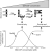
Model for the regulation of survival, proliferation and activation by the phosphotyrosine/phosphoserine binary switch. Shown is a schematic representation of a cytoplasmic portion of the βc subunit of the GM-CSF receptor encompassing Tyr577 and Ser585. Low concentrations of cytokine (<10 pM) promote PKA activation, Ser585 phosphorylation, 14-3-3 binding and PI 3-kinase recruitment. These signalling events are specifically linked to the regulation of cell survival only, and occur in the absence of detectable phosphotyrosine signalling pathways and cell proliferation/activation. Increasing the concentration of cytokine results in a switch in signalling whereby Tyr577 becomes phosphorylated, and blocks the phosphorylation of Ser585 by PKA. In addition, there is also a concomitant recruitment of phosphatases. This is accompanied by the binding of Shc to Tyr577, the phosphorylation of JAK2, STAT5 and ERK, and the regulation of both cell survival and cell proliferation/activation. In this model, Ser585 and Tyr577 function as a molecular switch that converts an analog input (GM-CSF concentration) to a binary output (either Ser585 signalling and survival, or Tyr577 signalling and survival together with proliferation/activation).

References

    1. Blume-Jensen P, Jiang G, Hyman R, Lee KF, O'Gorman S, Hunter T (2000) Kit/stem cell factor receptor-induced activation of phosphatidylinositol 3′-kinase is essential for male fertility. Nat Genet 24: 157–162 - PubMed
    1. Bulavin DV, Higashimoto Y, Demidenko ZN, Meek S, Graves P, Phillips C, Zhao H, Moody SA, Appella E, Piwnica-Worms H, Fornace AJ Jr (2003) Dual phosphorylation controls Cdc25 phosphatases and mitotic entry. Nat Cell Biol 5: 545–551 - PubMed
    1. Chen W, Yu YL, Lee SF, Chiang YJ, Chao JR, Huang JH, Chiong JH, Huang CJ, Lai MZ, Yang-Yen HF, Yen JJ (2001) CREB is one component of the binding complex of the Ces-2/E2A-HLF binding element and is an integral part of the interleukin-3 survival signal. Mol Cell Biol 21: 4636–4646 - PMC - PubMed
    1. Craparo A, Freund R, Gustafson TA (1997) 14-3-3 (ɛ) interacts with the insulin-like growth factor I receptor and insulin receptor substrate I in a phosphoserine-dependent manner. J Biol Chem 272: 11663–11669 - PubMed
    1. DeNichilo MO, Stewart AG, Vadas MA, Lopez AF (1991) Granulocyte-macrophage colony-stimulating factor is a stimulant of platelet-activating factor and superoxide anion generation by human neutrophils. J Biol Chem 266: 4896–4902 - PubMed

Publication types

MeSH terms

Substances