. 2006 Feb;80(4):1645-52.
doi: 10.1128/JVI.80.4.1645-1652.2006.

Generation of CD8+ T-cell responses by a recombinant nonpathogenic Mycobacterium smegmatis vaccine vector expressing human immunodeficiency virus type 1 Env

Affiliations

Generation of CD8+ T-cell responses by a recombinant nonpathogenic Mycobacterium smegmatis vaccine vector expressing human immunodeficiency virus type 1 Env

Mark J Cayabyab et al. J Virol. 2006 Feb.

Abstract

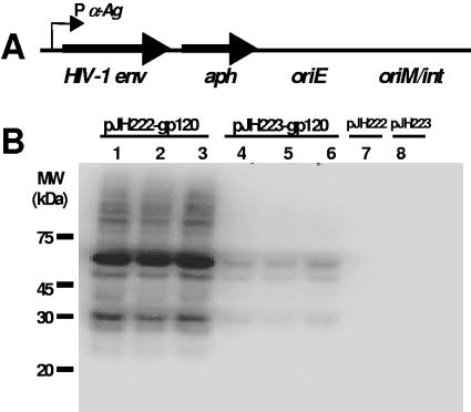
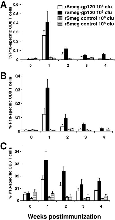
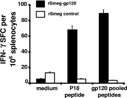
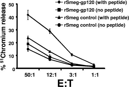
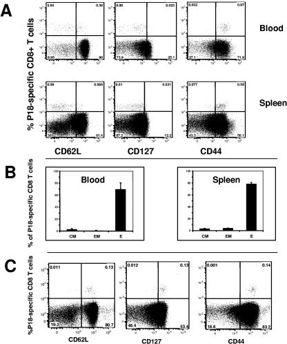
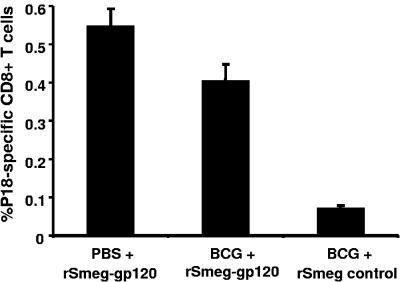
Because the vaccine vectors currently being evaluated in human populations all have significant limitations in their immunogenicity, novel vaccine strategies are needed for the elicitation of cell-mediated immunity. The nonpathogenic, rapidly growing mycobacterium Mycobacterium smegmatis was engineered as a vector expressing full-length human immunodeficiency virus type 1 (HIV-1) HXBc2 envelope protein. Immunization of mice with recombinant M. smegmatis led to the expansion of major histocompatibility complex class I-restricted HIV-1 epitope-specific CD8(+) T cells that were cytolytic and secreted gamma interferon. Effector and memory T lymphocytes were elicited, and repeated immunization generated a stable central memory pool of virus-specific cells. Importantly, preexisting immunity to Mycobacterium bovis BCG had only a marginal effect on the immunogenicity of recombinant M. smegmatis. This mycobacterium may therefore be a useful vaccine vector.


Figures

FIG. 1.
FIG. 1.
Expression of HIV-1 HXBc2 gp120 envelope in nonpathogenic Mycobacterium smegmatis. (A) Codon-optimized HXBc2 gp120 env was cloned into the E. coli/mycobacteria shuttle plasmids pJH222 (multicopy) and pJH223 (integrative). The gp120 env in the plasmids is under the control of the M. tuberculosis α-Ag promoter (P α-Ag). A fusion protein was created in which the M. tuberculosis 19-kDa signal sequence was at the N terminus and an influenza hemagglutinin (HA) epitope tag was at the C terminus of the gp120 Env. Both plasmids contained the Tn903-derived aph gene conferring kanamycin resistance as a selectable marker and an E. coli origin of replication (oriE). The origin of replication (oriM) was inserted into the pJH222 plasmid, while the attP site and the int gene of mycobacteriophage L5 were included in pJH223. (B) Western blot analysis showed expression of the gp120 protein in recombinant M. smegmatis MC2155. The gp120 expression of three independent clones of mycobacteria transformed with either pJH222-gp120 (lanes 1 to 3) or pJH223-gp120 (lanes 4 to 6) was determined using an anti-HA MAb (clone 3F10). Mycobacteria transformed with either mock pJH222 or pJH223 containing an irrelevant gene (malaria msp1) were utilized as negative controls (lanes 7 and 8). MW, molecular mass.
FIG. 2.
FIG. 2.
Recombinant M. smegmatis elicited HIV-1-specific CD8+ T-cell responses in mice. BALB/c mice were inoculated via the intraperitoneal route with approximately 106 CFU or 108 CFU gp120-expressing recombinant M. smegmatis (rSmeg-gp120) organisms transformed with either the integrative pJH223-gp120 plasmid (A) or multicopy pJH222-gp120 (B). As a negative control, mice were inoculated with the same dose of mycobacteria transformed with the control pJH222- and pJH223-msp1 plasmids (rSmeg control). (C) Mice were inoculated twice (10 weeks apart) with the same dose of either the rSmeg-gp120 (integrative) construct or the rSmeg control. The mean (± SEM) percent HIV-1 HXBc2 gp120 P18 tetramer-positive CD8 T cells from PBMC collected at the indicated time points is shown for each group of mice (n = 4 per group).
FIG. 3.
FIG. 3.
rM. smegmatis-elicited HIV-1-specific CD8+ T cells secreted IFN-γ. Day 7 splenocytes from mice immunized with 107 CFU recombinant mycobacteria expressing gp120 (integrative) were exposed to no peptide, P18, or a gp120 peptide pool and evaluated in an ELISPOT assay. Splenocytes from mice immunized with rM. smegmatis expressing Msp1 were used as a control. The mean (± SEM) spot-forming cells (SFC) per 106 splenocytes for each group of mice (n = 4 per group) is shown.
FIG. 4.
FIG. 4.
Cytotoxic activity of HIV-1-specific CD8+ T cells elicited by rM. smegmatis immunization. HIV-1-specific CTL were expanded in vitro by stimulating splenocytes isolated from mice at day 14 after a single inoculation with 108 CFU rM. smegmatis expressing gp120 (integrative) with 10 ng/ml p18 peptide in the presence of rat IL-2 for 7 days. Cytotoxic activity of the effector cells for P815 target cells pulsed with or without p18 was assessed in a 51chromium release assay. Effector-to-target (E:T) ratios used in the study are indicated.
FIG. 5.
FIG. 5.
Phenotype of HIV-1-specific CD8 T cells elicited by immunization with rM. smegmatis. Mice were immunized with 108 CFU rM. smegmatis expressing gp120 (integrative). (A) Flow cytometric analysis of week 1 PBMC and splenocytes from immunized mice revealed expression of CD44 on the surface of all rM. smegmatis-elicited tetramer-positive cells. CD62L and CD127 were expressed on a subset of the tetramer-positive cells. (B) The proportions of effector (P18-tetramer positive, CD127, and CD62Llo), effector memory (P18-tetramer positive, CD127+, and CD62Llo), and central memory (P18-tetramer positive, CD127+, and CD62Lhi) cells in the blood and spleen of mice immunized with rM. smegmatis are shown. Effector, effector memory, and central memory cells are denoted E, EM, and CM, respectively. The mean (± SEM) percent E, EM, or CM for each group of mice (n = 4 per group) is shown. (C) Peripheral blood HIV-1-specific CD8+ T cells from mice 1 year after immunization with rM. smegmatis expressing gp120 were predominantly central memory cells. PBMC were pooled from 4 mice that were inoculated twice (10 weeks apart) with 108 CFU bacilli.
FIG. 6.
FIG. 6.
Recombinant M. smegmatis-elicited HIV-1-specific CD8+ T-cell responses in mice preimmunized with BCG. Mice were immunized with wild-type BCG (Pasteur) or PBS and 6 months later with 108 CFU rM. smegmatis expressing gp120 (integrative) (indicated as BCG + rSmeg-gp120 and PBS + rSmeg-gp120, respectively). BCG-preimmunized mice that were subsequently inoculated with rSmeg control were used as a negative control (indicated as BCG + rSmeg control). Tetramer analysis was performed on the PBMC of mice 1 week after inoculation with the rM. smegmatis constructs. The mean (± SEM) percent of HIV-1 HXBc2 gp120 P18-tetramer-positive CD8+ T cells is shown for each group (n = 4 to 5 mice per group).

References

    1. Ahmad-Nejad, P., H. Hacker, M. Rutz, S. Bauer, R. M. Vabulas, and H. Wagner. 2002. Bacterial CpG-DNA and lipopolysaccharides activate Toll-like receptors at distinct cellular compartments. Eur. J. Immunol. 32:1958-1968. - PubMed
    1. Aldovini, A., and R. A. Young. 1991. Humoral and cell-mediated immune responses to live recombinant BCG-HIV vaccines. Nature 351:479-482. - PubMed
    1. Andersson, G. E., and P. M. Sharp. 1996. Codon usage in the Mycobacterium tuberculosis complex. Microbiology 142:915-925. - PubMed
    1. Appay, V., D. F. Nixon, S. M. Donahoe, G. M. Gillespie, T. Dong, A. King, G. S. Ogg, H. M. Spiegel, C. Conlon, C. A. Spina, D. V. Havlir, D. D. Richman, A. Waters, P. Easterbrook, A. J. McMichael, and S. L. Rowland-Jones. 2000. HIV-specific CD8(+) T cells produce antiviral cytokines but are impaired in cytolytic function. J. Exp. Med. 192:63-75. - PMC - PubMed
    1. Bange, F. C., F. M. Collins, and W. R. Jacobs, Jr. 1999. Survival of mice infected with Mycobacterium smegmatis containing large DNA fragments from Mycobacterium tuberculosis. Tuber. Lung Dis. 79:171-180. - PubMed

Publication types

LinkOut - more resources