. 2006 Mar 3;98(4):490-8.
doi: 10.1161/01.RES.0000205846.46812.be. Epub 2006 Jan 26.

High-density lipoprotein hydrolysis by endothelial lipase activates PPARalpha: a candidate mechanism for high-density lipoprotein-mediated repression of leukocyte adhesion

Affiliations

High-density lipoprotein hydrolysis by endothelial lipase activates PPARalpha: a candidate mechanism for high-density lipoprotein-mediated repression of leukocyte adhesion

Waleed Ahmed et al. Circ Res. .

Abstract

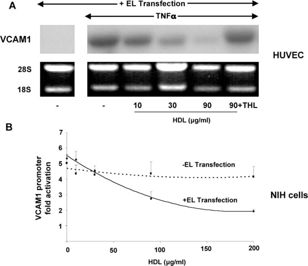
Although high-density lipoprotein (HDL) is known to inhibit endothelial adhesion molecule expression, the mechanism for this anti-inflammatory effect remains obscure. Surprisingly, we observed that HDL no longer decreased adhesion of U937 monocytoid cells to tumor necrosis factor (TNF)alpha-stimulated human endothelial cells (EC) in the presence of the general lipase inhibitor tetrahydrolipstatin. In considering endothelial mechanisms responsible for this effect, we found that endothelial lipase (EL) overexpression in both EC and non-EL-expressing NIH/3T3 mouse embryonic fibroblasts cells significantly decreased TNFalpha-induced VCAM1 expression and promoter activity in a manner dependent on HDL concentration and intact EL activity. Given recent evidence for lipolytic activation of peroxisome proliferator-activated receptors (PPARs)-nuclear receptors implicated in metabolism, atherosclerosis, and inflammation-we hypothesized HDL hydrolysis by EL is an endogenous endothelial mechanism for PPAR activation. In both EL-transfected NIH cells and bovine EC, HDL significantly increased PPAR ligand binding domain activation in the order PPAR-alpha> >-gamma>-delta. Moreover, HDL stimulation induced expression of the canonical PPARalpha-target gene acyl-CoA-oxidase (ACO) in a PPARalpha-dependent manner in ECs. Conditioned media from EL-adenovirus transfected cells but not control media exposed to HDL also activated PPARalpha. PPARalpha activation by EL was most potent with HDL as a substrate, with lesser effects on LDL and VLDL. Finally, HDL inhibited leukocyte adhesion to TNFalpha-stimulated ECs isolated from wild-type but not PPARalpha-deficient mice. This data establishes HDL hydrolysis by EL as a novel, distinct natural pathway for PPARalpha activation and identifies a potential mechanism for HDL-mediated repression of VCAM1 expression, with significant implications for both EL and PPARs in inflammation and vascular biology.


Figures

Figure 1
Figure 1
HDL inhibits adhesion of U937 monocytoid cells to TNFα-stimulated ECs but not in the presence of the lipase inhibitor tetrahydrolipstatin. A, Fluoresce-in-labeled U937 cells were added to TNFα-stimulated (50 ng/mL) human EC monolayers after pretreatment with the PPARα agonist WY14643 (10 µmol/L), HDL (90 µg/mL), or HDL (90 µg/mL) in the presence of the lipase inhibitor tetrahydrolipstatin (10 µmol/L). Fluorescent microscopy shows adherent U937 cells (green) on a near-confluent EC layer. B, Quantification of U937 adherence on EC monolayers as determined by fluorescence assay. Results are expressed as percentage of leukocytes bound to TNFα-stimulated cells. Bars represent mean±SEM (n=3); #P<0.01 TNFα-stimulated ECs vs control; *P<0.01 WY and HDL vs TNFα-stimulated ECs.
Figure 2
Figure 2
EL/HDL inhibits TNFα-mediated VCAM1 mRNA expression and promoter activity. A, Northern analysis of VCAM1 mRNA expression was performed on human EC (HUVEC) transfected with EL and then pretreated with increasing HDL concentrations (µg/mL, 3 hours) before TNFα (20 ng/mL, 16 hours) stimulation. In these EL-transfected cells, HDL decreased TNFα-induced VCAM1 expression but not in the presence of the lipase inhibitor tetrahydrolipstatin (10 µmol/L). B, TNFα- mediated VCAM1 promoter activation was tested in NIH cells over a concentration range of HDL in the presence (solid line) or absence (dashed line) of EL transfection. Cells were pretreated with HDL (3 hours) and then stimulated with TNFα (30 ng/mL, 12 hours) before performing promoterluciferase assays. In the presence of EL transfection, HDL significantly repressed VCAM1 promoter activation by 63% compared with 17% in EL nontransfected cells (*P<0.05 at 200 µg/mL of HDL; n=3, in triplicate for all). For comparison, the synthetic PPARα agonist WY14643 (250 µmol/L) suppressed TNFα-induced VCAM1 promoter activity from 5.3-fold to 1.7-fold; EL/HDL had no effect on the VCAM1 promoter in the presence of the lipase inhibitor tetrahydrolipstatin (10 µmol/L, data not shown).
Figure 3
Figure 3
EL preferentially hydrolyzes HDL to activate PPARα in a manner dependent on EL catalysis. Standard PPARα-LBD-GAL4 assays as described in Methods were performed in BAECs either with or without cotransfection of human EL. Results are expressed as fold activation over control after normalization. A, EL preferentially acts on phosphotidylcholine (PC) and HDL to activate the PPARα– LBD. EC transfected with either empty vector or the EL construct were exposed to PC, LDL, VLDL, and HDL at the concentrations shown and PPARα–LBD activity determined. In EL-transfected cells, the most potent activation was seen with HDL (*P<0.05); more modest activation was also evident with LDL and VLDL (all lipoproteins at 60 µg protein/mL). For comparison, the PPARα-LBD response to WY14643 (10 µmol/L) is shown. B, In untransfected BAEC, HDL increases PPARα-LBD activation in a concentration-dependent manner but not in the presence of tetrahydrolipstatin (10 µmol/L). C, Tetrahydrolipstatin preincubation (30 minutes, room temperature) decreases PPARα-LBD activation in a concentration-dependent manner as evident in control transfected (not shown) and EL-transfected BAEC exposed to HDL (60 µg/mL). D, PPARα activation by EL/HDL requires catalytically-active EL. PPARα–LBD-GAL4 assays were performed in BAEC after transfection of either wild-type or a catalytically-inactive EL point mutant in the presence of increasing concentrations of HDL. In cells expressing the catalytically-inactive EL mutant, no significant increase in PPARα–LBD activation is seen above basal levels across the HDL concentration tested. For comparison, concomitant PPARα-LBD activation in cells transfected with wild-type EL is shown; tetrahydrolipstatin (10 µmol/L) abrogated the EL/HDL/PPARα response.
Figure 4
Figure 4
Heterologous EL expression Confers PPARα Activation In Response to HDL. Standard PPARα–LBD-GAL4 assays were performed as before but in NIH cells known to lack endogenous EL expression. A, NIH cells transfected with either EL or the empty vector were exposed to increasing concentrations of HDL before PPARα–LBD activation was determined. HDL activates the PPARα–LBD in a concentration-dependent manner but only in EL transfected cells. B, PPARα–LBD assays were performed in NIH cells exposed to HDL (100 µg/mL) and increasing amounts of either EL protein conditioned medium (prepared and quantified from EL adenovirus-expressing cells as described in Methods) or control-transfected medium (50 µL/well to 500 µL/well of medium, 24-well plates). The extent of PPARα-LBD activation seen was linearly related to the amount of EL-conditioned medium added; control medium had no effect.
Figure 5
Figure 5
Hydrolysis of HDL by EL activates PPAR isotypes in the order -α>-γ >δ. The activation of each PPAR-LBD isotype (α, γ, or δ) in response to EL expression in the presence of increasing HDL concentrations was tested using LBD assays in NIH cells as before. EL/HDL most potently activated PPARα, followed by PPARγ, and then PPARδ at the maximal HDL concentration tested (90 µg/mL). The EL/HDL response was expressed as a percentage of the PPAR–LBD activation seen with known PPAR isotype-specific ligands as follows: PPARα-WY14643 (10 µmol/L); PPARγ-BRL49653 (1 µmol/L); PPARδ- bezafibrate (10 µmol/L).
Figure 6
Figure 6
EL/HDL induces mRNA expression of the PPARα target gene acyl CoA oxidase (ACO) but only in the genetic presence of PPARα. ECs from PPARα wild-type (+/+) and deficient (−/−) mice were transfected with EL or a control plasmid and stimulated with either WY14643 (10 µmol/L) or HDL (90 or 200 µg/mL) before performing Northern blot analysis for ACO. Densitometry and quantification analysis as compared with GAPDH expression indicates WY and HDL increased ACO 1.92-fold and 2.47-fold, respectively, in EL-transfected cells.
Figure 7
Figure 7
EL and LPL are distinct mechanisms for lipolytic PPARα activation. The additional PPARα–LBD activation seen with EL vs LPL in response to VLDL or HDL was compared in BAEC. A, Cells were exposed to increasing concentrations of LPL as shown in the presence of a fixed concentration of VLDL (5 µg/mL). As previously reported, LPL increased PPARα activation in a concentration-dependent manner. The presence or absence of EL-transfection had no impact on the PPAR activation seen by LPL/VLDL. B, Similar experiments as in (A) were repeated with increasing concentrations of LPL but in the presence of a fixed concentration of HDL (90 µg/mL). With HDL as the substrate, the presence of EL overexpression significantly increases the PPARα activation seen at any given LPL concentration (P<0.05 for each LPL concentration).
Figure 8
Figure 8
HDL inhibits leukocyte adhesion to TNFα-stimulated mouse ECs in a PPARα-dependent manner. A, Fluorescein-labeled murine J774A.1 mouse monocyte-macrophage-like cells were added to TNFα-stimulated (30 ng/mL) mouse PPARα+/+ and PPARα−/− ECs after pretreatment with either the PPARα agonist WY14643 (10 µmol/L), HDL (90 µg/mL), or HDL (90 µg/mL) in the presence of the lipase inhibitor tetrahydrolipstatin (10 µmol/L). Fluorescence microscopy shows adherent J774A.1 cells (green) on a confluent mouse EC layer. B, Quantification of J774A.1 adherence on mouse EC monolayers as determined by fluorescence assay. Results are expressed as percentage of leukocytes bound to TNFα-stimulated cells. Bars represent mean±SD (n=3); #P<0.05 (TNFα-stimulated alone vs TNFα-unstimulated control EC); **P<0.01 (TNFα-stimulated +/+ vs −/− ECs); *P<0.05 (TNFα and WY or HDL vs TNFα-stimulated alone +/+ ECs); †P<0.02 (unstimulated +/+ vs −/− ECs).

References

    1. Gordon T, Castelli WP, Hjortland MC, Kannel WB, Dawber TR. High density lipoprotein as a protective factor against coronary heart disease. The Framingham Study. Am J Med. 1977;62:707–714. - PubMed
    1. Castelli WP, Garrison RJ, Wilson PW, Abbott RD, Kalousdian S, Kannel WB. Incidence of coronary heart disease and lipoprotein cholesterol levels. The Framingham Study. JAMA. 1986;256:2835–2838. - PubMed
    1. Glass CK, Witztum JL. Atherosclerosis. the road ahead. Cel. 2001;104:503–516. - PubMed
    1. Cockerill GW, Rye KA, Gamble JR, Vadas MA, Barter PJ. High-density lipoproteins inhibit cytokine-induced expression of endothelial cell adhesion molecules. Arterioscler Thromb Vasc Biol. 1995;15:1987–1994. - PubMed
    1. Calabresi L, Franceschini G, Sirtori CR, De Palma A, Saresella M, Ferrante P, Taramelli D. Inhibition of VCAM-1 expression in endothelial cells by reconstituted high density lipoproteins. Biochem Biophys Res Commun. 1997;238:61–65. - PubMed

Publication types