

L1 is a potential marker for poorly-differentiated pancreatic neuroendocrine carcinoma
- PMID: 16440424
- PMCID: PMC4077503
- DOI: 10.3748/wjg.v12.i1.94
L1 is a potential marker for poorly-differentiated pancreatic neuroendocrine carcinoma
Abstract
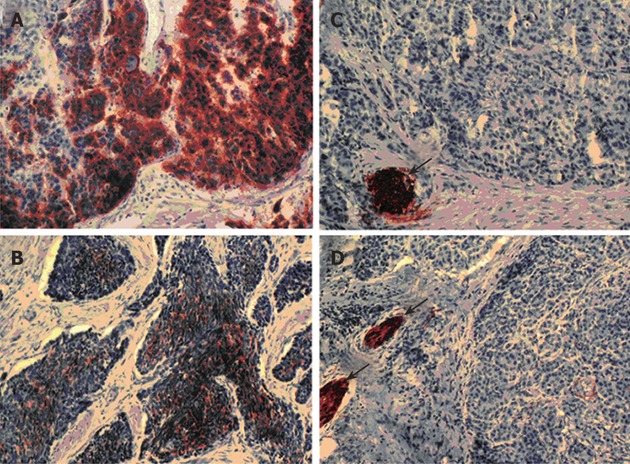
Aim: To determine the expression of L1 in pancreatic neuroendocrine tumor and to correlate it with WHO classification of this tumor.
Methods: We retrospectively analyzed L1 expression in 63 cases of pancreatic neuroendocrine tumor by immunohistochemistry on paraffin sections of primary tumors or metastases. Staining was performed by peroxidase technique with monoclonal antibody UJ127.11 against human L1. All tumors were classified according to WHO classification as well-differentiated neuroendocrine tumors and carcinomas or poorly-differentiated neuroendocrine carcinomas.
Results: L1 was detected in 5 (7.9%) of 63 pancreatic neuroendocrine tumors. Four (44.4%) of 9 poorly-differentiated carcinomas expressed L1. In contrast, only 1 (1.9%) of 54 well-differentiated tumors or carcinomas was positive for L1. No expression was found in Langerhans islet cells of normal pancreatic tissue. Cross table analysis showed a significant association between L1 expression and classification of neuroendocrine tumors of the pancreas (P<0.01).
Conclusion: L1 is specifically expressed in poorly-differentiated pancreatic neuroendocrine carcinomas that are known to have the worst prognosis. L1 might be a marker for risk prediction of patients diagnosed with pancreatic neuroendocrine carcinomas.
Figures
References
-
- Rindi G, Klöppel G. Endocrine tumors of the gut and pancreas tumor biology and classification. Neuroendocrinology. 2004;80 Suppl 1:12–15. - PubMed
-
- Rindi G, Villanacci V, Ubiali A. Biological and molecular aspects of gastroenteropancreatic neuroendocrine tumors. Digestion. 2000;62 Suppl 1:19–26. - PubMed
-
- Heymann MF, Joubert M, Nemeth J, Franc B, Visset J, Hamy A, le Borgne J, le Neel JC, Murat A, Cordel S, et al. Prognostic and immunohistochemical validation of the capella classification of pancreatic neuroendocrine tumours: an analysis of 82 sporadic cases. Histopathology. 2000;36:421–432. - PubMed
-
- Solcia E, Kloppel G, Sobin LH. Histological typing of endocrine tumors. In: World Health Organization International Histological Classification of Endocrine Tumors., editor. 2 ed. New York: Springer; 2000. pp. 38–74.
-
- Klöppel G, Perren A, Heitz PU. The gastroenteropancreatic neuroendocrine cell system and its tumors: the WHO classification. Ann N Y Acad Sci. 2004;1014:13–27. - PubMed
Publication types
MeSH terms
Substances
LinkOut - more resources
Full Text Sources
Other Literature Sources
Medical
