

Clinical utility of quantitative RT-PCR targeted to alpha1,4-N-acetylglucosaminyltransferase mRNA for detection of pancreatic cancer
- PMID: 16441422
- PMCID: PMC11158275
- DOI: 10.1111/j.1349-7006.2006.00148.x
Clinical utility of quantitative RT-PCR targeted to alpha1,4-N-acetylglucosaminyltransferase mRNA for detection of pancreatic cancer
Erratum in
- Cancer Sci. 2006 Mar;97(3):242
Abstract
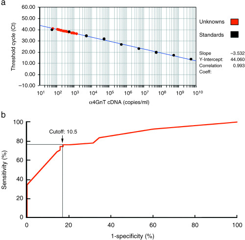
alpha1,4-N-Acetylglucosaminyltransferase (alpha4GnT) is a glycosyltransferase responsible for the biosynthesis of alpha1,4-GlcNAc-capped O-glycans, and is frequently expressed in pancreatic cancer cells but not peripheral blood cells. In the present study, we tested the clinical utility of alpha4GnT mRNA expressed in the mononuclear cell fraction of peripheral blood as a biomarker of pancreatic cancer. Total RNA isolated from the peripheral blood mononuclear cells from 55 pancreatic cancer patients, 10 chronic pancreatitis patients, and 70 cancer-free volunteers was analyzed quantitatively by reverse transcription-polymerase chain reaction with primers specific for alpha4GnT, and the expression level of alpha4GnT mRNA relative to that of glyceraldehyde-3-phosphate dehydrogenase (GAPDH) was measured. When the ratio of alpha4GnT to GAPDH transcripts exceeded a defined cut-off value, patients were considered to have pancreatic cancer. By these standards, 76.4% of the pancreatic cancer patients were detected by this assay. A strong correlation was obtained between positivity in this assay and the expression of alpha4GnT protein detected immunohistochemically in pancreatic cancer tissues resected subsequently, suggesting that alpha4GnT mRNA detected in the peripheral blood is derived from circulating pancreatic cancer cells. Although increased levels of alpha4GnT mRNA was detected in 40.0% of chronic pancreatitis patients and 17.1% of cancer-free volunteers, the expression levels were significantly lower than those seen in pancreatic cancer patients. These results suggest that quantitative analysis of alpha4GnT mRNA expressed in the mononuclear cell fraction of peripheral blood will contribute to the detection of pancreatic cancer.
Figures


Similar articles
-
Usefulness of the real-time reverse transcription-polymerase chain reaction assay targeted to alpha1,4-N-acetylglucosaminyltransferase for the detection of gastric cancer.Lab Invest. 2003 Feb;83(2):187-97. doi: 10.1097/01.lab.0000057001.21187.a0. Lab Invest. 2003. PMID: 12594234
-
Glycosylation of MUC6 by α1,4-linked N-acetylglucosamine enhances suppression of pancreatic cancer malignancy.Cancer Sci. 2022 Feb;113(2):576-586. doi: 10.1111/cas.15209. Epub 2021 Nov 28. Cancer Sci. 2022. PMID: 34808019 Free PMC article.
-
Expression cloning of a human alpha1, 4-N-acetylglucosaminyltransferase that forms GlcNAcalpha1-->4Galbeta-->R, a glycan specifically expressed in the gastric gland mucous cell-type mucin.Proc Natl Acad Sci U S A. 1999 Aug 3;96(16):8991-6. doi: 10.1073/pnas.96.16.8991. Proc Natl Acad Sci U S A. 1999. PMID: 10430883 Free PMC article.
-
Expression of mesothelin mRNA in pure pancreatic juice from patients with pancreatic carcinoma, intraductal papillary mucinous neoplasm of the pancreas, and chronic pancreatitis.Pancreas. 2005 May;30(4):349-54. doi: 10.1097/01.mpa.0000160281.56828.76. Pancreas. 2005. PMID: 15841046
-
[Glycosyltransferase genes as tumor marker].Rinsho Byori. 2002 Nov;Suppl 123:142-8. Rinsho Byori. 2002. PMID: 12652802 Review. Japanese.
Cited by
-
A Comprehensive Review of the Potential Role of Liquid Biopsy as a Diagnostic, Prognostic, and Predictive Biomarker in Pancreatic Ductal Adenocarcinoma.Cells. 2023 Dec 19;13(1):3. doi: 10.3390/cells13010003. Cells. 2023. PMID: 38201207 Free PMC article. Review.
-
Liquid biopsy in patients with pancreatic cancer: Circulating tumor cells and cell-free nucleic acids.World J Gastroenterol. 2016 Jul 7;22(25):5627-41. doi: 10.3748/wjg.v22.i25.5627. World J Gastroenterol. 2016. PMID: 27433079 Free PMC article. Review.
-
Diagnostic and prognostic biomarkers in pancreatic carcinoma.Int J Clin Exp Pathol. 2009;2(1):1-10. Epub 2008 Apr 12. Int J Clin Exp Pathol. 2009. PMID: 18830385 Free PMC article.
-
Circulating RNAs as new biomarkers for detecting pancreatic cancer.World J Gastroenterol. 2015 Jul 28;21(28):8527-40. doi: 10.3748/wjg.v21.i28.8527. World J Gastroenterol. 2015. PMID: 26229396 Free PMC article. Review.
-
Analysis of microRNAs in pancreatic fine-needle aspirates can classify benign and malignant tissues.Clin Chem. 2008 Oct;54(10):1716-24. doi: 10.1373/clinchem.2008.109603. Epub 2008 Aug 21. Clin Chem. 2008. PMID: 18719196 Free PMC article.
References
Publication types
MeSH terms
Substances
Grants and funding
LinkOut - more resources
Full Text Sources
Medical
Molecular Biology Databases
Research Materials
