. 2006 Feb;97(2):139-47.
doi: 10.1111/j.1349-7006.2006.00144.x.

1alpha,25-Dihydroxyvitamin D3 downmodulates the functional differentiation of Th1 cytokine-conditioned bone marrow-derived dendritic cells beneficial for cytotoxic T lymphocyte generation

Affiliations

1alpha,25-Dihydroxyvitamin D3 downmodulates the functional differentiation of Th1 cytokine-conditioned bone marrow-derived dendritic cells beneficial for cytotoxic T lymphocyte generation

Junko Matsuzaki et al. Cancer Sci. 2006 Feb.

Abstract

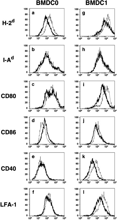
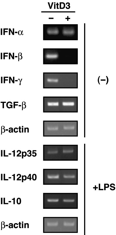
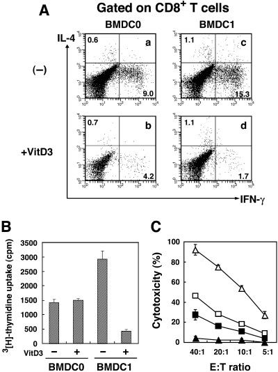
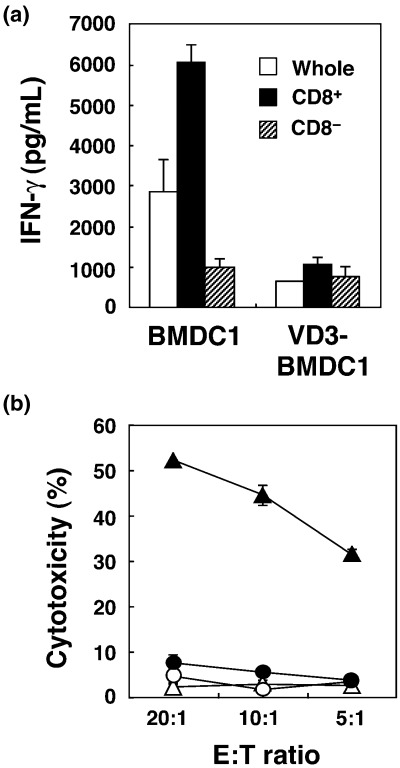
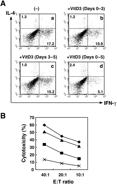
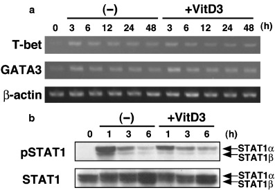
Various dendritic cell subsets are induced from bone marrow cells under different cytokine conditions. We have demonstrated previously that the Th1-cytokine-conditioned bone marrow-derived dendritic cell (BMDC) subset BMDC1 (generated in the presence of granulocyte-macrophage colony-stimulating factor [GM-CSF] + interleukin [IL]-3 + interferon [IFN]-gamma+ IL-12) induces a much stronger type 1 immune response than BMDC0 (GM-CSF + IL-3). In the present study, we investigated the effect of 1alpha,25-dihydroxyvitamine D3 (VitD3), which is a known immunomodulating drug, on the differentiation of BMDC subsets. The addition of VitD3 significantly influenced the functional differentiation of BMDC1 compared with BMDC0. Specifically, the addition of VitD3 greatly decreased the expression levels of MHC class I, CD80, CD40 and leukocyte function-associated antigen (LFA)-1 molecules on BMDC1. In addition, VitD3-treated BMDC1 (VD3-BMDC1) almost completely lost their immunostimulating activity for inducing type 1 immunity and cytotoxic T lymphocyte generation. A failure in the induction of type 1 immunity by VD3-BMDC1 appeared to be due to the following: (i) the expression of co-stimulatory molecules on VD3-BMDC1 was strongly downmodulated compared with BMDC1 generated without VitD3; and (ii) VD3-BMDC1 showed significantly lower mRNA expression of IFN-gamma and IFN-beta, factors that are essential for cytotoxic T lymphocyte induction. VitD3 inhibited the differentiation of functionally competent BMDC1 during the early phase of differentiation but not during the late differentiation period. A possible reason for the inhibition of BMDC1 differentiation by VitD3 is reduced phosphorylation of STAT1 during early differentiation. Taken together, VitD3 strongly suppressed T-cell responses by inhibiting functional differentiation of precursor dendritic cells into functional BMDC1 that are feasible for inducing Th1-dependent cellular immunity.


Figures

Figure 1
Figure 1
The expression of functional cell surface molecules on BMDC1 was greatly downmodulated by VitD3. (a–f) BMDC0 and (g–l) BMDC1 were generated from bone marrow cells by culture under distinct culture conditions with or without VitD3. Expression of cell surface molecules ([a,g] H‐2d, [b,h] I‐Ad, [c,i] CD80, [d,j] CD86, [e,k] CD40 and [f,l] leukocyte function‐associated antigen [LFA]‐1) was determined by flow cytometry. The data indicate the staining profile of BMDC subsets generated in the presence (–––) or absence (. . .) of VitD3. Similar results were obtained in three different experiments.
Figure 2
Figure 2
VitD3 inhibited mRNA expression of interferon (IFN)‐β and IFN‐γ in the BMDC1 subset. The mRNA expression of cytokines (IFN‐α, IFN‐β, IFN‐γ and transforming growth factor (TGF)‐β) in BMDC1 and VD3‐BMDC1 was determined by reverse transcription–polymerase chain reaction. After lipopolysaccharide (LPS) stimulation for 48 h, the expression of interleukin (IL)‐12p35, IL‐12p40 and IL‐10 mRNA was analyzed. To exclude mRNA expression by other cells types, CD11c+ cells were purified by cell sorting before extraction of total RNA. Similar results were obtained in three different experiments.
Figure 3
Figure 3
VitD3 reduced the capability of BMDC1 to induce alloantigen‐specific cytotoxic T lymphocytes (CTL) in a mixed lymphocyte reaction (MLR). Spleen cells of C57BL/6 were stimulated with mytomicin C (MMC)‐treated allogeneic BMDC0 or BMDC1 generated with or without VitD3 for 4 days. (A) The production of interleukin (IL)‐4 and interferon (IFN)‐γ from CD8+ T cells stimulated with BMDC0 (a,b) or BMDC1 (c,d) generated in the absence (a,c) or in the presence (b,d) of VitD3 was determined by intracellular cytokine staining. Numbers represent the percentage of cells in each quadrant. The same results were obtained in three separate experiments. (B) After 4 days of MLR, cells were pulsed with 3[H]‐thymidine for 4 h and harvested on a glass filter. Proliferation of spleen cells stimulated with BMDC subsets was determined by counting incorporated 3H using a β‐counter. The bars represent the mean ± SE of triplicate samples. (C) Alloantigen‐specific CTL activity of C57BL/6 mouse spleen cells was measured after 4 day culture with BALB/c BMDC subsets generated in the presence or absence of VitD3. After 4 days of MLR, cells were harvested and their cytotoxicity against P815 mastocytoma cells (H‐2d) was measured using a 4‐h 51Cr‐release assay. The bars represent the mean ± SE of triplicate samples. □, BMDC0; ▵, BMDC1; ▪, VD3‐BMDC0; ▴, VD3‐BMDC1.
Figure 4
Figure 4
BMDC1 generated with VitD3 failed to generate cytotoxic T lymphocytes (CTL) from isolated CD8+ T cells. CD8+ or CD8 cells separated from spleen cells of C57BL/6 mice were stimulated with BMDC1 or VD3‐BMDC1 induced from bone marrow cells of BALB/c mice. (a) Interferon (IFN)‐γ levels of the supernatant of culture at day 4 were determined by ELISA. The bars represent the mean ± SE of triplicate samples. (b) Alloantigen‐specific CTL activity was measured using a 4‐h 51Cr‐release assay against P815 mastocytoma cells. The bars represent the mean ± SE of triplicate samples. ▴, CD8+ (BMDC1); ▵, CD8 (BMDC1); •, CD8+ (VD3‐BMDC1); ○, CD8 (VD3‐BMDC1).
Figure 5
Figure 5
The inhibitory effect of VitD3 was exhibited during the early stage but not the late stage of BMDC1 differentiation. BMDC1 were generated from bone marrow cells in the absence or presence of VitD3. VitD3 was added during days 0–3, days 3–5 or days 0–5 (VD3‐BMDC1). (A) CD8+ T cells stimulated with allogeneic BMDC1 for 4 days were further activated by plate‐bound anti‐CD3 monoclonal antibody for 6 h, and cytoplasmic interleukin‐4 and interferon‐γ production were assessed by flow cytometry. (B) Cytotoxic T lymphocyte activity of alloantigen‐specific T cells against P815 cells (H‐2d) was measured using a 4‐h 51Cr‐release assay. Bars represent the mean ± SE from three separate experiments. ◆, (–); ▪, +VitD3 (days 0–3); ▴, +VitD3 (days 3–5); ×, +VitD3 (days 0–5).
Figure 6
Figure 6
VitD3 inhibited phosphorylation of STAT1 at the early stage of BMDC1 differentiation. CD4CD8B220CD11bGr‐1 precursor bone marrow cells were stimulated under BMDC1 conditions with or without VitD3. (a) T‐bet and GATA‐3 mRNA expression was analyzed by reverse transcription–polymerase chain reaction at different times. As an internal control, expression of β‐actin was also analyzed. (b) Expression of phospho‐STAT1 and STAT1 was determined by western blot analysis.

Similar articles

Cited by

References

    1. Banchereau J, Steinman RB. Dendritic cells and the control of immunity. Nature 1998; 392: 245–52. - PubMed
    1. Palucka K, Banchereau J. Dendritic cells: a link between innate and adaptive immunity. J Clin Immunol 1999; 19: 12–25. - PubMed
    1. Sato M, Iwakabe K, Ohta A et al. Functional heterogeneity among bone marrow‐derived dendritic cells conditioned by Th1‐ and Th2‐biasing cytokines for the generation of allogeneic cytotoxic T lymphocytes. Int Immunol 2000; 12: 335–42. - PubMed
    1. Sato M, Chamoto K, Nishimura T. A novel tumor‐vaccine cell therapy using bone marrow‐derived dendritic cell type 1 and antigen‐specific Th1 cells. Int Immunol 2003; 15: 837–43. - PubMed
    1. Steinbrink K, Wolfl M, Jonuleit H, Knop J, Enk AH. Induction of tolerance by IL‐10‐treated dendritic cells. J Immunol 1997; 159: 4772–80. - PubMed

MeSH terms