Comparative Study
. 2006 Mar;92(3):234-41.
doi: 10.1016/j.ijgo.2005.12.001. Epub 2006 Jan 25.

Measurement of the pubic portion of the levator ani muscle in women with unilateral defects in 3-D models from MR images

Affiliations
Comparative Study

Measurement of the pubic portion of the levator ani muscle in women with unilateral defects in 3-D models from MR images

L Chen et al. Int J Gynaecol Obstet. 2006 Mar.

Abstract

Objective: Develop a method to quantify the cross-sectional area of the pubic portion of the levator ani muscle, validate the method in women with unilateral muscle defects, and report preliminary findings in those women.

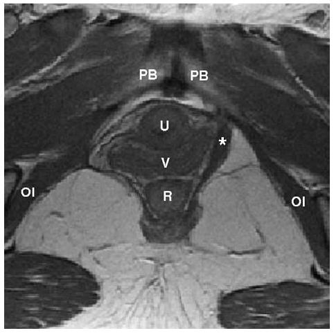
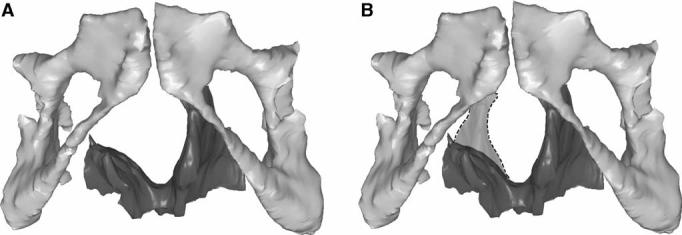
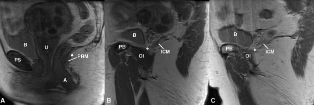
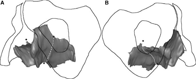
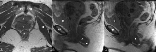
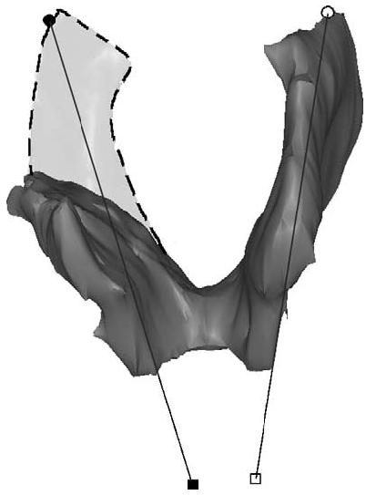
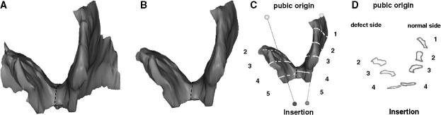
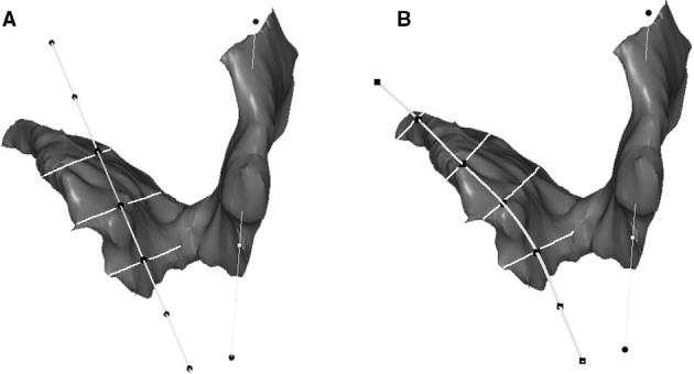
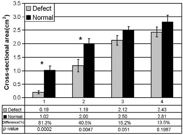
Method: Multi-planar proton density magnetic resonance images of 12 women with a unilateral defect in the pubic portion of their levator ani were selected from a larger study of levator ani muscle anatomy in women with and without genital prolapse. Three-dimensional bilateral models of the levator ani were reconstructed (using 3-D Slicer, version 2.1b1) and divided into iliococcygeal and pubic portions. Muscle cross-sectional areas were calculated at four equally spaced locations perpendicular to a line drawn from the pubic origin to the visceral insertion using the I-DEAS computer modeling software.

Results: The cross-sectional area of the muscle on the side with the defect was smaller than the normal side at all the four locations. The average bilateral difference was up to 81% at location 1 (nearest pubic origin). Almost all of the volume difference (13.7%, P=0.0004) was attributable to a reduction in the pubic portion (24.6%, P<0.0001), not the iliococcygeal portion (P=0.64), of the muscle.

Conclusions: A method was developed to quantify cross-sectional area of the pubic portion of the levator ani perpendicular to the intact muscle direction. Significant bilateral cross-sectional area differences were found between intact and defective muscles in women with a unilateral defect.


Figures

Figure 1
Figure 1
Axial proton density MR image of a woman with a right unilateral defect. The left levator ani muscle is intact (denoted by *) while the right side portion of the muscle is missing. PB denotes pubic bone; U: urethra; V: vagina; R: rectum; OI: obturator internus muscle.
Figure 2
Figure 2
(A) Reconstructed 3-D model showing the pubic bone and levator ani muscle with a right unilateral defect. (B) The dashed region shows the expected location of the missing muscle after reflecting the muscle from the normal side across the midline.
Figure 3
Figure 3
Separating the pubic portion of levator ani from the iliococcygeus portion on MR scans. A): Identification of the middle point (triangle) on the mid-sagittal MRI; B) and C): identification of the two lateral points on MRI (cross and diamond). The following structures were identified in MRI to help for orientation. PS denotes pubic symphysis; PB: pubic bone; B: bladder; A: anus; OI: obturator internus; U: uterus (with fibroids); PRM: puborectalis muscle. ICM: iliococcygeus muscle.
Figure 4
Figure 4
3-D view of the separation of the pubic portion from the iliococcygeal portion of the levator ani. The dashed line represents the plane of separation, while the triangle, cross and diamond have the same meaning as in the preceding figure.
Figure 5
Figure 5
Reference points for determining fiber direction in the pubic portion of the levator ani. A) Origin points on axial MR at its junction with obturator internus (OI) as open circle on patient's left and filled circle on patient's right; B) and C) right (filled square) and left (open square) insertion point on sagittal MR scans. The following structures are identified aid orientation. PB: pubic bone; B: bladder; V: vagina; R: rectum; A: anus.
Figure 6
Figure 6
Lines connecting the origin and insertion points (see preceding figure) were used to estimate muscle fiber direction. The dashed region shows the expected location of the muscle on the defective side, were it intact.
Figure 7
Figure 7
Methodological steps to quantify levator ani muscle loss in a subject with a unilateral defect. A: Frontal view of the 3-D reconstruction model of levator ani was built. B: The pubic portion is separated from the iliococcygeal portion; here only the pubic portion is shown. C: The line of estimated fiber direction of the pubic portion was determined. Equally spaced cutting planes perpendicular to the fiber direction line were placed. D: The resulting cross-sections are shown. Cross-sectional areas were then calculated. Due to the defect on the right, muscle is not present at location 1; therefore, it is not shown.
Figure 8
Figure 8
A: Cross-sectional area perpendicular to ‘normal’ muscle direction; B: Cross-sectional area perpendicular to remaining muscle direction.
Figure 9
Figure 9
Bilateral comparison of muscle cross-sectional area of pubic portion of levator ani muscle perpendicular to its fiber direction. Bars are standard error and * denotes P <0.05.

References

    1. Olsen AL, Smith VJ, Bergstrom JO, Colling JC, Clark AL. Epidemiology of surgically managed pelvic organ prolapse and urinary incontinence. Obstet Gynecol. 1997;89:501–6. - PubMed
    1. Boreham MK, Wai CY, Miller RT, Schaffer JL, Word RA. Morphometric analysis of smooth muscle in the anterior vaginal wall of women with pelvic organ prolapse. Am J Obstet Gynecol. 2002;187:56–63. - PubMed
    1. Boreham MK, Wai CY, Miller RT, Schaffer JL, Word RA. Morphometric properties of the posterior vaginal wall in women with pelvic organ prolapse. Am J Obstet Gynecol. 2002;187:1501–9. - PubMed
    1. Takano CC, Girao MJ, Sartori MG, Castro RA, Arruda RM, Simoes MJ, et al. Analysis of collagen in parametrium and vaginal apex of women with and without uterine prolapse. Int Urogynecol J. 2002;13(6):342–5. - PubMed
    1. Smith ARD, Hosker GL, Warrell DW. The role of partial denervation of the pelvic floor in the etiology of genitourinary prolapse and stress incontinence of urine: a neurophysiologic study. Br J Obstet Gynaecol. 1989;96:24–8. - PubMed

Publication types