

Review
doi: 10.1016/j.mce.2005.12.049.
Epub 2006 Jan 27.
Estrogen mediated cross talk between the ovary and pituitary somatotrope. Pre-ovulatory support for reproductive activity
Affiliations
- PMID: 16443322
- PMCID: PMC1751516
- DOI: 10.1016/j.mce.2005.12.049
Item in Clipboard
Review
Estrogen mediated cross talk between the ovary and pituitary somatotrope. Pre-ovulatory support for reproductive activity
Mol Cell Endocrinol.
.
No abstract available
Figures
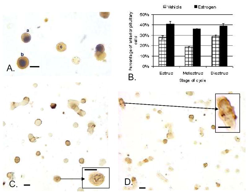
A. Dual labeling for Estrogen Receptor –Alpha isoform (ERα) in the nucleus (black) and growth hormone in the cytoplasm (orange-amber) of freshly dispersed cells from diestrous female rat. Most GH cells in the field express ERα. Cell C does not. B.Exposure to estradiol for 24 h (100 pM) stimulated expression of biotinylated Growth hormone releasing hormone (Bio-GHRH) binding sites, if given to estrous, metestrous, or diestrous female rats. C. Illustration of Bio-GHRH binding (gray-black) and labeling for growth hormone (orange-amber) in vehicle-treated diestrous female rats. D. Increased labeling for both Bio-GHRH (black) and GH (orange) is seen after 24 h in 100 pM estradiol. Insets show a higher magnification of dual labeled GH cells. Bar=15 μm
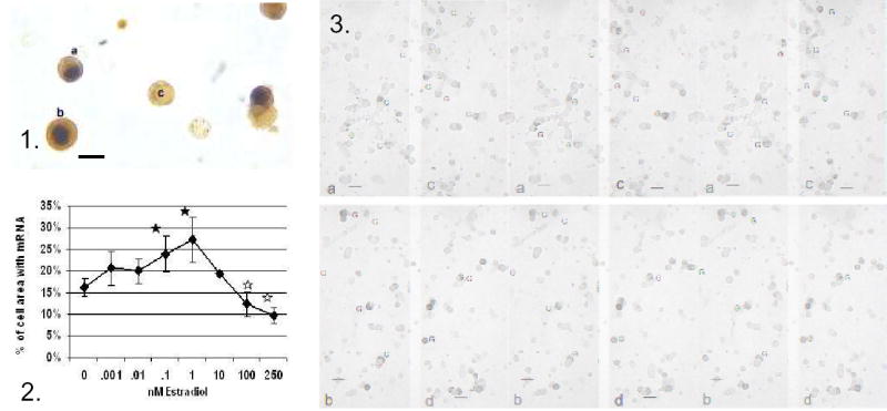
Figure 1.. Dual labeling for Estrogen Receptor –Alpha isoform (ERα) in the nucleus (black) and growth hormone in the cytoplasm (orange-amber) of freshly dispersed cells from diestrous female rat. Most GH cells in the field express ERα. Figure 2. Image analysis showed that 24 h in 0.1–1 nM estradiol produced an increase in average area of label for GH mRNA. Concentrations higher than 10 nM, however produced a decrease in labeling for the mRNA. Figure 3. shows this change by illustrating labeling for GH mRNA in cultures treated with vehicle (a), or 0.1 nM (b), 1 nM (c), or 250 nM estradiol. Bar=15 μm
Similar articles
-
Thyroid hormone and reproduction: regulation of estrogen receptors in goldfish gonads.Mol Reprod Dev. 2010 Sep;77(9):784-94. doi: 10.1002/mrd.21219. Mol Reprod Dev. 2010. PMID: 20722048
-
Management of epilepsy throughout the reproductive cycle--an overview of treatment issues.Axone. 1999 Sep;21(1):18-20. Axone. 1999. PMID: 10732520 Review. No abstract available.
-
Growth hormone-releasing hormone and extra-pituitary tumorigenesis: therapeutic and diagnostic applications of growth hormone-releasing hormone antagonists.Expert Opin Investig Drugs. 2003 Aug;12(8):1385-94. doi: 10.1517/13543784.12.8.1385. Expert Opin Investig Drugs. 2003. PMID: 12882623 Review.
-
Estrogen receptors are involved in xenoestrogen induction of growth hormone in the rat pituitary gland.J Reprod Dev. 2009 Apr;55(2):206-13. doi: 10.1262/jrd.20147. Epub 2009 Jan 15. J Reprod Dev. 2009. PMID: 19145065
-
Luteinizing hormone-releasing factor potentiates lordosis behavior in hypophysectomized ovariectomized female rats.Science. 1973 Dec 14;182(4117):1148-9. doi: 10.1126/science.182.4117.1148. Science. 1973. PMID: 4584371
Cited by
-
Ablation of leptin signaling to somatotropes: changes in metabolic factors that cause obesity.Endocrinology. 2012 Oct;153(10):4705-15. doi: 10.1210/en.2012-1331. Epub 2012 Aug 3. Endocrinology. 2012. PMID: 22865370 Free PMC article.
-
Effects of genistein on stereological and hormonal characteristics of the pituitary somatotrophs in rats.Endocrine. 2014 Dec;47(3):869-77. doi: 10.1007/s12020-014-0265-3. Epub 2014 Apr 22. Endocrine. 2014. PMID: 24752394
-
Ghrelin restoration of function in vitro in somatotropes from male mice lacking the Janus kinase (JAK)-binding site of the leptin receptor.Endocrinology. 2013 Apr;154(4):1565-76. doi: 10.1210/en.2012-2254. Epub 2013 Feb 15. Endocrinology. 2013. PMID: 23417423 Free PMC article.
-
Paracrinicity: the story of 30 years of cellular pituitary crosstalk.J Neuroendocrinol. 2008 Jan;20(1):1-70. doi: 10.1111/j.1365-2826.2007.01616.x. J Neuroendocrinol. 2008. PMID: 18081553 Free PMC article. Review.
-
The somatotrope as a metabolic sensor: deletion of leptin receptors causes obesity.Endocrinology. 2011 Jan;152(1):69-81. doi: 10.1210/en.2010-0498. Epub 2010 Nov 17. Endocrinology. 2011. PMID: 21084451 Free PMC article.
References
-
- Bethea CL. Estrogen action on growth hormone in pituitary cell cultures from adult and juvenile macaques. Endocrinology. 1991;129:2110–2118. - PubMed
-
- Chaidarun SS, Klibanski A, Alexander JM. Tumor-specific expression of alternatively spliced estrogen receptor messenger ribonucleic acid variants in human pituitary adenomas. J Clin Endocrinol Metab. 1997;82:1058–1065. - PubMed
-
- Chaidarun SS, Swearingen B, Alexander JM. Differential expression of estrogen receptor-β (ERβ) in human pituitary tumors: functional interactions with ERα and a tumor-specific splice variant. J Clin Endocrinol Metab. 1998;83:3308–3315. - PubMed
-
- Childs GV, Naor Z, Hazum E, Tibolt R, Westlund KM, Hancock MB. Localization of biotinylated gonadotropin releasing hormone on pituitary monolayer cells with avidin–biotin peroxidase complexes. J Histochem Cytochem. 1983a;31:1422–1425. - PubMed
-
- Childs GV, Naor Z, Hazum E, Tibolt R, Westlund KN, Hancock MB. Cytochemical characterization of pituitary target cells for biotinylated gonadotropin releasing hormone. Peptides. 1983b;4(4):549–555. - PubMed
Publication types
MeSH terms
Substances
Grants and funding
LinkOut - more resources
Full Text Sources
