. 2006 Feb 7;103(6):1888-93.
doi: 10.1073/pnas.0507493103. Epub 2006 Jan 27.

Small-molecule MDM2 antagonists reveal aberrant p53 signaling in cancer: implications for therapy

Affiliations

Small-molecule MDM2 antagonists reveal aberrant p53 signaling in cancer: implications for therapy

Christian Tovar et al. Proc Natl Acad Sci U S A. .

Abstract

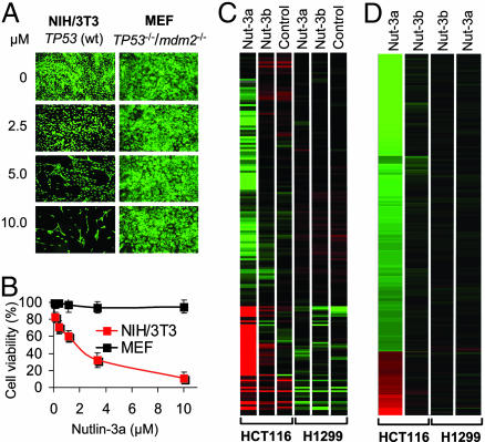
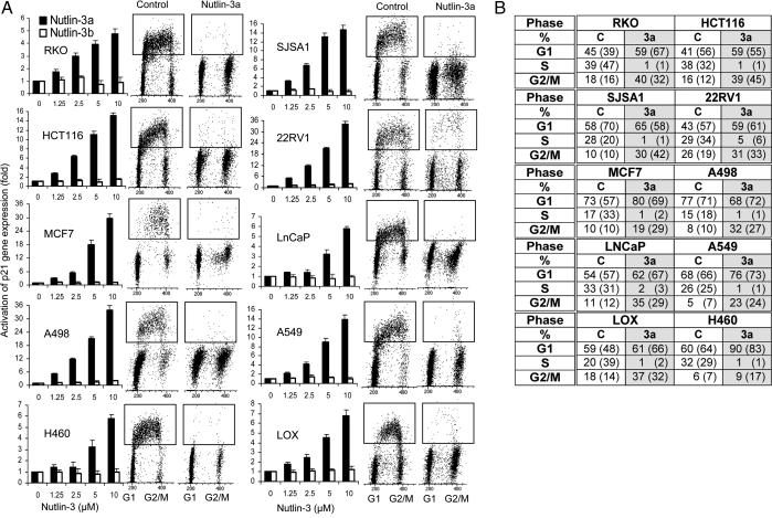
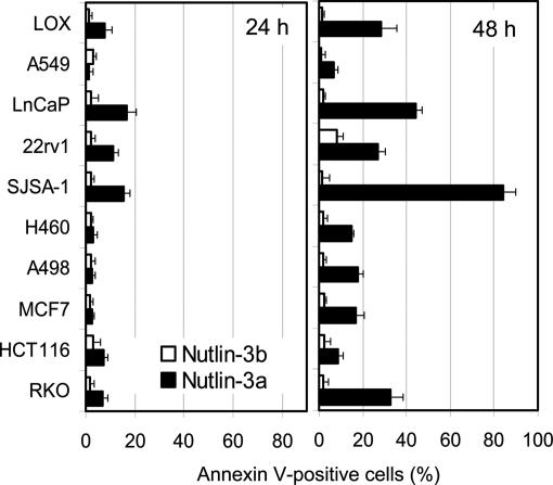
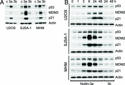
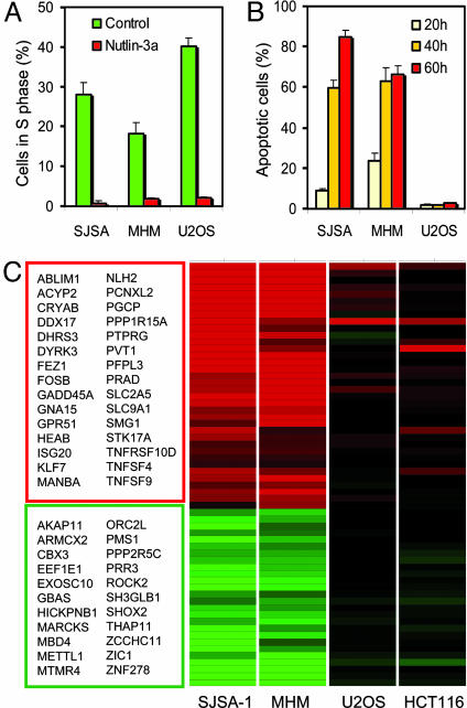
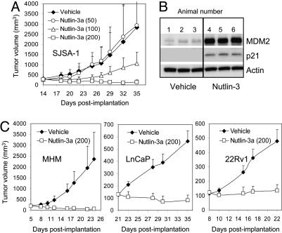
The p53 tumor suppressor retains its wild-type conformation and transcriptional activity in half of all human tumors, and its activation may offer a therapeutic benefit. However, p53 function could be compromised by defective signaling in the p53 pathway. Using a small-molecule MDM2 antagonist, nutlin-3, to probe downstream p53 signaling we find that the cell-cycle arrest function of the p53 pathway is preserved in multiple tumor-derived cell lines expressing wild-type p53, but many have a reduced ability to undergo p53-dependent apoptosis. Gene array analysis revealed attenuated expression of multiple apoptosis-related genes. Cancer cells with mdm2 gene amplification were most sensitive to nutlin-3 in vitro and in vivo, suggesting that MDM2 overexpression may be the only abnormality in the p53 pathway of these cells. Nutlin-3 also showed good efficacy against tumors with normal MDM2 expression, suggesting that many of the patients with wild-type p53 tumors may benefit from antagonists of the p53-MDM2 interaction.


Conflict of interest statement

Conflict of interest statement: No conflicts declared.

Figures

Fig. 1.
Fig. 1.
Nutlin-3 selectively activates p53. (A) Exponentially growing mouse NIH/3T3 fibroblasts (wt-TP53) and MEFs from TP53−/−/mdm2−/− knockout mice were incubated for 7 days in the presence of nutlin-3a and stained live by acridine orange. (B) Exponentially growing cells were treated with nutlin-3a for 5 days, and their viability was measured in three independent samples by the 3-(4,5-dimethylthiazol-2-yl)-2,5-diphenyl tetrazolium bromide assay and expressed as percentage of control ± SD. (C) A heat map of probe sets differentially expressed between nutlin-3a and all other treatments (3-fold changed, t test P < 0.05 vs. untreated cells) represented as the median normalized expression of the condition. Red indicates higher than median expression; green indicates lower than median expression. Data were generated on Affymetrix HG-U133 Genechips. (D) A heat map of the same probe sets displayed as a ratio of the treated condition vs. the untreated control.
Fig. 2.
Fig. 2.
Nutlin-activated p53 induces G1 and G2 arrest in cancer cell lines with wild-type p53. (A) Human cancer lines in exponential growth were treated with nutlin-3a or nutlin-3b for 24 h, and the expression of the p53 target gene p21 was measured by quantitative real-time PCR and expressed as fold increase ± SD. Cell-cycle profile before and after treatment with 10 μM nutlin-3a or an equivalent amount of solvent for 24 h was analyzed by BrdUrd labeling and flow cytometry. (B) Cell-cycle distribution was calculated from the flow cytograms in A and expressed as the percentage of the total population rounded to the full percentage value. Cell-cycle distribution from an independent experiment is shown in parentheses.
Fig. 3.
Fig. 3.
Apoptotic response of cancer cells with wild-type p53 to nutlin-3. Cells in exponential phase were treated with 10 μM nutlin-3a or nutlin-3b for 24 or 48 h, and the percentage of Annexin V-positive cells (live plus dead) was determined by the Guava Nexin kit and expressed as a percentage of the total cell number as in ref. . Data bars represent the average of two independent experiments.
Fig. 4.
Fig. 4.
Nutlin-3 stabilizes p53 and induces the p53 pathway in osteosarcoma cells. (A) Exponentially proliferating SJSA-1, MHM, and U2OS cells were treated with 10 μM nutlin-3a, nutlin-3b, or DMSO (c) for 24 h, and p53, p21, and MDM2 proteins in the cell lysates were analyzed by Western blotting. (B) Cells were treated with 10 μM nutlin-3a or nutlin-3b for the indicated times and analyzed by Western blotting. Representative results from two independent experiments are shown.
Fig. 5.
Fig. 5.
Nutlin-3-induced gene profiles differ between cells with high and low apoptotic index. (A) Nutlin-3a effectively arrests cell-cycle progression in the osteosarcoma cell lines SJSA-1, MHM, and U2OS. Cell-cycle analysis was performed as in Fig. 2. (B) Apoptosis induced by nutlin-3a in osteosarcoma cells. Treatment with nutlin-3a and determination of Annexin V-positive cell fraction was as in Fig. 3. (C) Differentially expressed genes in cells with high and low apoptotic index after 24 h incubation with 10 μM nutlin-3a. Expression profile was determined by Affymetrix gene array analysis (HG-U95 GeneChips) in cells treated with nutlin-3a or nutlin-3b. A heat map of the 62 probe sets found to be differentially expressed in SJSA-1 and MHM cells, but not in U2OS or HCT116, is shown. Differential expression was determined by a 3-fold change compared with untreated controls and an ANOVA P < 0.001 under a simple model including treatment, cell line, and their interaction. Red indicates activated genes; green indicates inhibited genes.
Fig. 6.
Fig. 6.
In vivo activity of nutlin-3a. (A) Nude mice bearing established SJSA-1 tumor xenografts received nutlin-3a for 3 weeks, and tumor growth was followed periodically. Error bars show SD from the mean volume (P < 0.01 for 200 mg/kg). (B) Nude mice with established SJSA-1 xenografts (three animals per group) received three oral doses of nutlin-3a (150 mg/kg) or vehicle and were killed 3 h after the last dose. Tumors were harvested and analyzed for the presence of the p53 targets MDM2 and p21 by Western blotting. (C) Nude mice with established tumor xenografts (100–200 mm3) were treated orally twice daily with 200 mg/kg nutlin-3a for 2 weeks (LnCaP and 22Rv1) or 3 weeks (MHM), and tumor volumes were recorded periodically. Error bars show SD from the mean volume. P was < 0.01 for all treatments.

Comment in

  • Protein-protein interactions for cancer therapy.
    Harris CC. Harris CC. Proc Natl Acad Sci U S A. 2006 Feb 7;103(6):1659-60. doi: 10.1073/pnas.0510948103. Epub 2006 Feb 1. Proc Natl Acad Sci U S A. 2006. PMID: 16452164 Free PMC article. No abstract available.

References

    1. Levine A. J. Cell. 1997;88:323–331. - PubMed
    1. Vogelstein B., Lane D., Levine A. J. Nature. 2000;408:307–310. - PubMed
    1. Harris S. L., Levine A. J. Oncogene. 2005;24:2899–2908. - PubMed
    1. Hollstein M., Sidransky D., Vogelstein B., Harris C. C. Science. 1991;253:49–53. - PubMed
    1. Hainaut P., Hollstein M. Adv. Cancer Res. 2000;77:81–137. - PubMed

MeSH terms