. 2006 Feb 7;103(6):1924-9.
doi: 10.1073/pnas.0510655103. Epub 2006 Jan 30.

The diacylglycerol-binding protein alpha1-chimaerin regulates dendritic morphology

Affiliations

The diacylglycerol-binding protein alpha1-chimaerin regulates dendritic morphology

Philip Buttery et al. Proc Natl Acad Sci U S A. .

Abstract

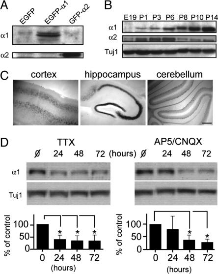
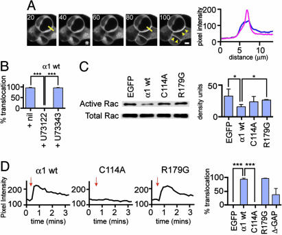
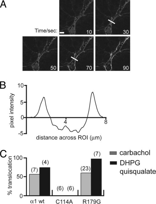
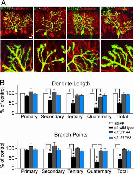
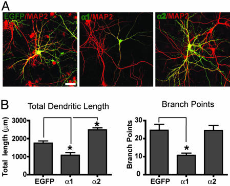
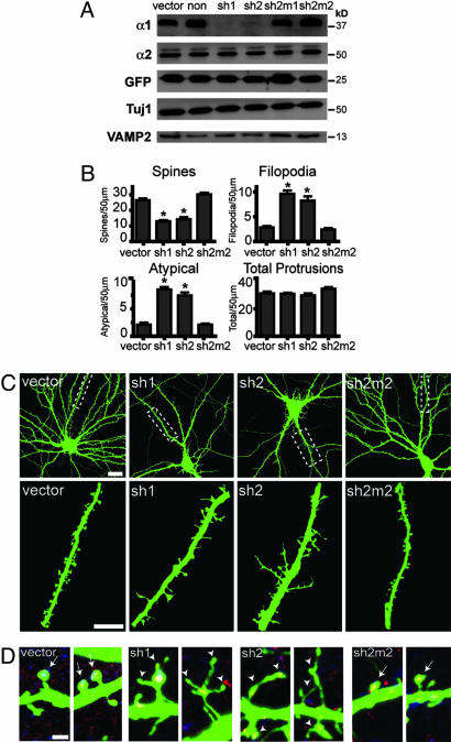
The morphological and functional differentiation of neuronal dendrites is controlled through transcriptional programs and cell-cell signaling. Synaptic activity is thought to play an important role in the maturation of dendritic arbors, but the signaling pathways that couple neuronal activity and morphological changes in dendrites are not well understood. We explored the function of alpha1-chimaerin, a neuronal diacylglycerol-binding protein with a Rho GTPase-activating protein domain that inactivates Rac1. We find that stimulation of phospholipase Cbeta-coupled cell surface receptors recruits alpha1-chimaerin to the plasma membrane of cultured hippocampal neurons. We further show that alpha1-chimaerin protein levels are controlled by synaptic activity and that increased alpha1-chimaerin expression results in the pruning of dendritic spines and branches. This pruning activity requires both the diacylglycerol-binding and Rac GTPase-activating protein activity of alpha1-chimaerin. Suppression of alpha1-chimaerin expression resulted in increased process growth from the dendritic shaft and from spine heads. Our data suggest that alpha1-chimaerin is an activity-regulated Rho GTPase regulator that is activated by phospholipase Cbeta-coupled cell surface receptors and contributes to pruning of dendritic arbors.


Conflict of interest statement

Conflict of interest statement: No conflicts declared.

Figures

Fig. 1.
Fig. 1.
Expression of α1-chimaerin. (A) Lysates from HEK 293 cells expressing EGFP, EGFP-α1-chimaerin, or EGFP-α2-chimaerin were probed with α1- and α2-specific antibodies. (B) Lysates from rat hippocampal tissue [embryonic day 19 (E19) through postnatal day 14 (P14)] were analyzed with α-chimaerin isoform-specific antibodies. (C) In situ hybridization on cortical, hippocampal, and cerebellar mouse tissue at postnatal day 10. No labeling was seen with sense probes (not shown). (Scale bar, 200 μm.) (D) (Upper) Western blot analysis with α1-specific antibodies on lysates from hippocampal cultures exposed to either tetrodotoxin (TTX) (1.5 μM), or 2-amino-5-phosphonopentanoic acid (AP5, 50 μM) and 6-cyano-7-nitroquinoxaline-2,3-dione (CNQX, 10 μM). (Lower) Relative protein levels were quantified by densitometric scanning (n = 3; ∗, P < 0.05).
Fig. 2.
Fig. 2.
EGFP-α1-chimaerin recruitment in response to DAG signaling. (A) (Left) Time-lapse microscopy of HEK 293 cells expressing mAChR1 and EGFP-α1-chimaerin-R179G. The asterisk marks time of stimulation with 5 mM carbachol. (Right) Intensity plot through cell junction (yellow line), before (blue) and 40 sec after stimulation (pink). (Scale bar, 5 μm.) (B) The percentage of cells exhibiting translocation was scored for control conditions compared with presence of either U73122 (20 μM) or U73343 (20 μM). The result is the mean of three independent experiments; ∗∗∗, P < 0.0005. (C) Point mutations in the C1 (C114A) and GAP (R179G) domains of α1-chimaerin result in decreased Rac-GAP activity as measured by PAK1 CRIB-domain pull-down assays in HEK 293 cells. Active and total Rac1 were detected by Western blotting (Left), and quantitated (Right) (n = 3; ∗, P < 0.05). (D) (Left) Translocation of wild-type and mutant EGFP-α1-chimaerins shown as the peak fluorescence intensity at the site of cell–cell junctions. Frames were captured every 10 sec, and red arrows mark the addition of carbachol. (Right) Percentage of cells exhibiting translocation (∗, P < 0.05).
Fig. 3.
Fig. 3.
Plasma membrane translocation of α1-chimaerin in hippocampal neurons. (A) Frames from a representative time-lapse series of a hippocampal neuron transfected with EGFP-α1-chimaerin-R179G and stimulated with carbachol (5 mM). Frames are shown after digital subtraction from the first frame. (B) Line scan of fluorescence intensity change from frame acquired at 30 and 70 sec (white line in A). (C) Percentage of cells exhibiting translocation to the plasma membrane upon stimulation with carbachol (5 mM, gray bars) or with the metabotropic glutamate receptor (mGluR) agonists (S)-3,5-dihydroxyphenylglycine (DHPG) or quisqualate (200 μM and 250 μM, respectively, black bars). Numbers in parentheses give total number of observations.
Fig. 4.
Fig. 4.
Effect of α1-chimaerin on dendritic arbors and dendritic spines. (A) Purkinje cells in cerebellar slices were transfected with EGFP and wild-type or each of the two mutant EGFP-α1-chimaerins. All Purkinje cells are labeled for EGFP (green) and calbindin (red). (Scale bars, 20 μm.) (B) Quantitation of total dendritic length (Upper) and dendritic branch points (Lower) in wild-type and mutant α1-chimaerin-expressing Purkinje cells (n > 10 cells; ∗, P > 0.05).
Fig. 5.
Fig. 5.
Overexpression of α-chimaerins in dissociated hippocampal neurons. (A) Dissociated hippocampal neurons were transfected at 10 days in vitro with EGFP, EGFP-α1-chimaerin, or EGFP-α2-chimaerin and labeled with EGFP (green) and microtubule-associated protein 2 (red) antibodies. (Scale bar, 40 μm.) (B) Quantitation of total dendritic length and number of branch points (n > 10 cells; ∗, P < 0.05).
Fig. 6.
Fig. 6.
Suppression of α1-chimaerin in hippocampal neurons results in excess outgrowth of dendritic protrusions. (A) Dissociated hippocampal neurons were infected with lentiviral vectors encoding shRNAs targeted against α1-chimaerin (sh1 and sh2), with mutated shRNAs (sh2m1 and sh2m2), with lentiviruses lacking an shRNA insert (vector), or with an active control hairpin (“non,” against p53). Lysates were probed with antibodies against α1-chimaerin, α2-chimaerin, Tuj1, and VAMP2. Comparable viral infection was confirmed by probing for EGFP, which was coexpressed with the shRNAs. (B and C) Morphometric analysis (B) and representative images (C) from hippocampal neurons transfected with control, sh1, sh2, and sh2m2 shRNA vectors (n > 10 cells; ∗, P < 0.05). (Scale bars, 20 μm and 10 μm for the whole-cell images and the enlarged region, respectively.) (D) Spine phenotypes from control and knockdown cells. Control and sh2m2 cells exhibited normal stubby and mushroom-headed spines (arrows); whereas, sh1 and sh2 cells show an increase in atypical spines with filopodia emanating from the spine head (arrowheads). PSD-95 (red) and VGLUT1 (blue) staining visualize synaptic terminals.

Similar articles

Cited by

References

    1. Bonhoeffer T., Yuste R. Neuron. 2002;35:1019–1027. - PubMed
    1. Wong R. O., Ghosh A. Nat. Rev. Neurosci. 2002;3:803–812. - PubMed
    1. Niell C. M., Meyer M. P., Smith S. J. Nat. Neurosci. 2004;7:254–260. - PubMed
    1. Sin W. C., Haas K., Ruthazer E. S., Cline H. T. Nature. 2002;419:475–480. - PubMed
    1. Hua J. Y., Smear M. C., Baier H., Smith S. J. Nature. 2005;434:1022–1026. - PubMed

Publication types