. 2006 Feb 7;103(6):1953-8.
doi: 10.1073/pnas.0509386103. Epub 2006 Jan 30.

Abeta and tau form soluble complexes that may promote self aggregation of both into the insoluble forms observed in Alzheimer's disease

Affiliations

Abeta and tau form soluble complexes that may promote self aggregation of both into the insoluble forms observed in Alzheimer's disease

Jian-Ping Guo et al. Proc Natl Acad Sci U S A. .

Abstract

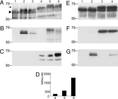
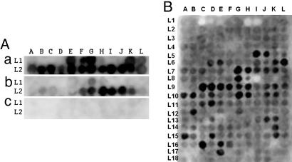
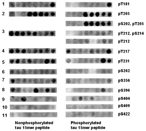
To date, there is no reasonable explanation as to why plaques and tangles simultaneously accumulate in Alzheimer's disease (AD). We demonstrate here by Western blotting and ELISA that a stable complex can form between tau and amyloid-beta protein (Abeta). This complex enhances tau phosphorylation by GSK3beta, but the phosphorylation then promotes dissociation of the complex. We have localized the sites of this interaction by using peptide membrane arrays. Abeta binds to multiple tau peptides, especially those in exons 7 and 9. This binding is sharply reduced or abolished by phosphorylation of specific serine and threonine residues. Conversely, tau binds to multiple Abeta peptides in the mid to C-terminal regions of Abeta. This binding is also significantly decreased by GSK3beta phosphorylation of tau. We used surface plasmon resonance to determine the binding affinity of Abeta for tau and found it to be in the low nanomolar range and almost 1,000-fold higher than tau for itself. In soluble extracts from AD and control brain tissue, we detected Abeta bound to tau in ELISAs. We also found by double immunostaining of AD brain tissue that phosphorylated tau and Abeta form separate insoluble complexes within the same neurons and their processes. We hypothesize that in AD, an initial step in the pathogenesis may be the intracellular binding of soluble Abeta to soluble nonphosphorylated tau, thus promoting tau phosphorylation and Abeta nucleation. Blocking the sites where Abeta initially binds to tau might arrest the simultaneous formation of plaques and tangles in AD.


Conflict of interest statement

Conflict of interest statement: No conflicts declared.

Figures

Fig. 1.
Fig. 1.
Western blotting of tau–Aβ complexes. (A–C) Western blotting after incubation of T40 only (lane 1), T40 plus Aβ40 (lane 2), T40 plus Aβ42 (lane 3), T40 plus GSK-3β (lane 4), T40 plus Aβ40 with GSK-3β (lane 5), and T40 plus Aβ42 with GSK-3β (lane 6) probed with antibodies Tau 12 (A), 4G8 (B), or PHF-1 (C). In A and B, the 30 μg/ml T40, 75 μg/ml Aβ40 and Aβ42, and 300 μg/ml GSK3β were used. The solutions were diluted 1:5 with SDS sample buffer, and 20 μl was loaded in each lane. In C and D, 5 μg/ml T40, 2.7 μg/ml Αβ40 and Aβ42, and 62.5 μg/ml GSK3β were used. The solutions were diluted 1:5 with SDS sample buffer, and 20 μl was loaded in each lane. In the samples of lanes 5 and 6, GSK-3β was added after incubation of T40 with Aβ40 and Aβ42. (D) Densitometric analysis of the bands in C. Each experiment was replicated twice with similar results. EG illustrate the negative effect of tau phosphorylation on the formation of tau–Aβ binding. Phosphorylation of T40 by GSK-3β (300 μg/ml) preceded coincubation of T40 (25 μg/ml) and Aβ40 (62.5 μg/ml). Blots were probed with A0024 (E), PHF-1 (F), and tAβ40 (G). Lane 1, T40; lane 2, T40 plus Aβ40; lane 3, GSK-3β-treated T40; lane 4, GSK-3β-treated T40 plus Aβ40. Samples were diluted 1:5 with SDS sample buffer, and 20 μl was loaded in each lane. Molecular weight markers are shown on the left. See Results for interpretation of the bands.
Fig. 2.
Fig. 2.
Interaction of recombinant T40 and phosphorylated T40 on Aβ 10-mer peptide array membranes, and Aβ1-42 on Tau 15-mer peptide array membrane. (A) 10 μg/ml recombinant T40 (a) or phosphorylated T40 by GSK-3β (b), or vehicle solution only (c), in 50 mM ammonium acetate (pH 6.5), 3 μM heparin (MW 6000), and 0.1 mM PMSF solution probed on Aβ 10-mer peptide array membrane, developed with ECL reagent after incubating with tau-2 monoclonal antibody and HRP–anti-mouse IgG conjugate (a and c), tau phosphorylation-dependent antibody pT231, and HRP–anti-rabbit IgG conjugate (b). (B) 10 μg/ml of Aβ1-42 in PBS probed on Tau 15-mer peptide array membrane, developed with ECL reagent after incubating with 4G8 monoclonal antibody and HRP–anti-mouse IgG conjugate.
Fig. 3.
Fig. 3.
Comparison of Aβ1-42 probing on 11 regions of tau where serine and threonine residues were either phosphorylated or not phosphorylated. The 15-mer peptide arrays were probed with Aβ1-42 (10 μg/ml in PBS). They were developed with ECL reagent after incubating with 4G8 monoclonal antibody and HRP–anti-mouse IgG conjugate.
Fig. 4.
Fig. 4.
Coexpression of Aβ and tau in neurofibrillary tangles. (AC) Tau (A0024 labeled with Hilyte Fluor 488) exhibiting a green fluorescence (A) and Aβ (4G8 labeled with Hilyte Fluor 555) showing red fluorescence (B) are coexpressed in tangle bearing neurons of the entorhinal cortex in a case of AD as shown by the yellow fluorescence in the merged image (C). (D) A lower-power merged image showing a mixture of tangled neurons coexpressing Aβ and tau (yellow color) and tau only (green color) as well as extracellular Aβ deposits (red color). (Scale bars, 50 μm A and 100 μm in D.)

References

    1. Iqbal K., Alonso Adel C., Chen S., Cholan M. O., El-Akkad E., Gong C. X., Khatoon S., Li B., Liu F., Rahman A., et al. Biochim. Biophys. Acta. 2005;1739:198–210. - PubMed
    1. Hardy J. A., Higgins G. A. Science. 1992;256:184–185. - PubMed
    1. Hardy J., Selkoe D. J. Science. 2002;297:353–356. - PubMed
    1. Hutton M. Movement Disorders. 2002;17:1402–1403.
    1. Goedert M., Jakes R. Biochim. Biophys. Acta. 2005;1739:240–250. - PubMed

Publication types

MeSH terms