

First molecular and biochemical analysis of in vivo affinity maturation in an ectothermic vertebrate
- PMID: 16446445
- PMCID: PMC1413636
- DOI: 10.1073/pnas.0508341103
First molecular and biochemical analysis of in vivo affinity maturation in an ectothermic vertebrate
Erratum in
- Proc Natl Acad Sci U S A. 2006 Apr 4;103(14):5632
Abstract
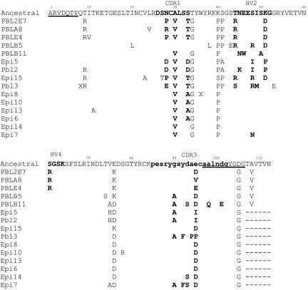
The cartilaginous fish are the oldest phylogenetic group in which Igs have been found. Sharks produce a unique Ig isotype, IgNAR, a heavy-chain homodimer that does not associate with light chains. Instead, the variable (V) regions of IgNAR bind antigen as soluble single domains. Our group has shown that IgNAR plays an integral part in the humoral response of nurse sharks (Ginglymostoma cirratum) upon antigen challenge. Here, we generated phage-displayed libraries of IgNAR V regions from an immunized animal and found a family of clones derived from the same rearrangement event but differentially mutated during expansion. Because of the cluster organization of shark Ig genes and the paucicopy nature of IgNAR, we were able to construct the putative ancestor of this family. By studying mutations in the context of clone affinities, we found evidence that affinity maturation occurs for this isotype. Subsequently, we were able to identify mutations important in the affinity improvement of this family. Because the family clones were all obtained after immunization, they provide insight into the in vivo maturation mechanisms, in general, and for single-domain antibody fragments.
Conflict of interest statement
Conflict of interest statement: No conflicts declared.
Figures


References
-
- Eisen H. N., Siskind G. W. Biochemistry. 1964;155:996–1008. - PubMed
-
- Griffiths G. M., Berek C., Kaartinen M., Milstein C. Nature. 1984;312:271–275. - PubMed
-
- Berek C., Griffiths G. M., Milstein C. Nature. 1985;316:412–418. - PubMed
-
- Berek C., Berger A., Apel M. Cell. 1991;67:1121–1129. - PubMed
-
- Ziegner M., Steinhauser G., Berek C. Eur. J. Immunol. 1994;24:2393–2400. - PubMed
Publication types
MeSH terms
Substances
Associated data
- Actions
- Actions
- Actions
- Actions
- Actions
- Actions
- Actions
- Actions
- Actions
- Actions
- Actions
- Actions
- Actions
- Actions
- Actions
- Actions
- Actions
- Actions
- Actions
Grants and funding
LinkOut - more resources
Full Text Sources
Other Literature Sources
