. 2006 Feb 7;103(6):1768-73.
doi: 10.1073/pnas.0510452103. Epub 2006 Jan 30.

Calorie restriction induces mitochondrial biogenesis and bioenergetic efficiency

Affiliations

Calorie restriction induces mitochondrial biogenesis and bioenergetic efficiency

G López-Lluch et al. Proc Natl Acad Sci U S A. .

Abstract

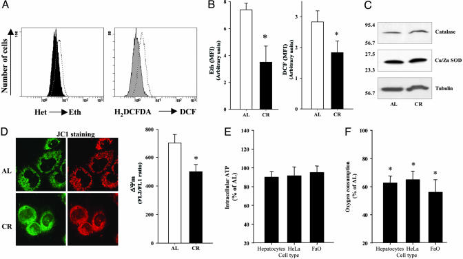
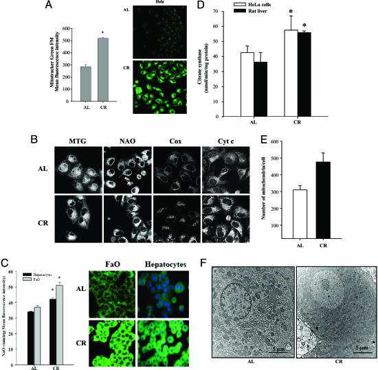
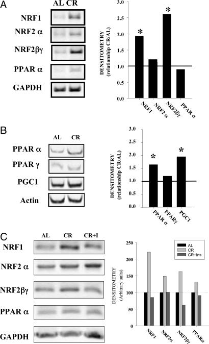
Age-related accumulation of cellular damage and death has been linked to oxidative stress. Calorie restriction (CR) is the most robust, nongenetic intervention that increases lifespan and reduces the rate of aging in a variety of species. Mechanisms responsible for the antiaging effects of CR remain uncertain, but reduction of oxidative stress within mitochondria remains a major focus of research. CR is hypothesized to decrease mitochondrial electron flow and proton leaks to attenuate damage caused by reactive oxygen species. We have focused our research on a related, but different, antiaging mechanism of CR. Specifically, using both in vivo and in vitro analyses, we report that CR reduces oxidative stress at the same time that it stimulates the proliferation of mitochondria through a peroxisome proliferation-activated receptor coactivator 1 alpha signaling pathway. Moreover, mitochondria under CR conditions show less oxygen consumption, reduce membrane potential, and generate less reactive oxygen species than controls, but remarkably they are able to maintain their critical ATP production. In effect, CR can induce a peroxisome proliferation-activated receptor coactivator 1 alpha-dependent increase in mitochondria capable of efficient and balanced bioenergetics to reduce oxidative stress and attenuate age-dependent endogenous oxidative damage.


Conflict of interest statement

Conflict of interest statement: No conflicts declared.

Figures

Fig. 1.
Fig. 1.
ROS in HeLa cells cultured under AL and CR serum conditions. Oxidation of Het or cH2DCFDA in cells cultured after 48 h with AL or CR sera was determined separately by flow cytometry as indicated in Methods. (A) Histogram distribution from a significant experiment of each determination. Dotted histogram corresponds to AL cells, and filled histogram corresponds to CR cells. Eth, ethidium; DCF, dichlorofluorescein. (B) Data indicate the mean of the mean fluorescence intensity (MFI) from three different experiments performed in duplicate. *, Significant differences vs. AL treatment, P < 0.01. (C) Protein levels of catalase and Cu/Zn SOD. Western blots were performed as described in Methods. (D) ΔΨm in HeLa cells cultured under AL and CR conditions after 48 h was determined by using JC1 as indicated in Methods. (Left) Confocal images of JC1-stained cells after treatment. Images were acquired by using a ×63 objective. (Right) Ratiometric analysis of the fluorescence of JC1-aggregated form (FL2) vs. JC1-free form (FL1) from three different experiments performed in duplicate. *, Significant differences vs. AL treatment, P < 0.01. (E and F) Intracellular ATP levels (E) and oxygen consumption (F) in cells after 48 h of incubation. *, Significant differences vs. AL oxygen consumption, P < 0.01.
Fig. 2.
Fig. 2.
Mitochondrial mass and distribution in HeLa cells cultured under AL and CR serum conditions. (A) Mitochondrial mass in cells cultured after 48 h with AL or CR sera was determined by flow cytometry by using MTG as indicated in Methods. (Left) Data indicate the mean of the MFI from three different experiments performed in duplicate. *, Significant differences vs. AL treatment, P < 0.01. (Right) MTG signal in AL and CR cells visualized by confocal microscopy. Images were acquired by using a ×40 objective. Settings for detectors were maintained along the study. (B) Mitochondrial staining in HeLa cells cultured under AL and CR serum conditions. Representative images of mitochondrial staining in AL and CR cells by using 20 nM MTG, 10 μM NAO, and immunostaining with anticytochrome c (Cyt c) or anticytochrome c oxidase I (Cox). Images were acquired by using a ×63 objective. (C) NAO staining of FAO and primary rat hepatocytes cultured in AL and CR conditions. (Left) Data indicate the mean of the MFI from three different experiments performed in duplicate. *, Significant differences vs. AL treatment, P < 0.01. (Right) NAO signal in AL and CR cells visualized by confocal microscopy. Images were acquired by using the ×40 objective. (D) Citrate synthase activity in HeLa cells incubated with rat serum grown under AL or CR conditions and from rat liver from animals fed under these conditions for 8 months. *, Significant differences vs. AL treatment, P < 0.01. (E) Quantification of the mitochondrial numbers from micrographs of 24-month-old AL and CR rat hepatocytes as described in Methods. *, Significant differences vs. AL treatment, P < 0.01. (F) EM of AL (Left) and CR (Right) rat hepatocytes prepared and imaged as described in Methods.
Fig. 3.
Fig. 3.
Regulation of mitochondrial biogenesis. (A) Expression of mitochondrial biogenesis factors determined by RT-PCR in HeLa cells. Total RNA from cells cultured during 48 h with 10 AL or CR sera was extracted, and the amount of specific mitochondrial factors was determined by RT-PCR as indicated in Methods. The average intensity of each product was related to the control gene GAPDH or β-actin. These ratios were then used to calculate relative mRNA levels by densitometry. (Left) A representative image of blots from three different experiments is shown. (Right) Data represent the mean of the relationship between the mRNA from CR samples and AL samples from at least three different experiments. *, Significant increase of blots in CR samples vs. AL samples, P < 0.05. (B) Expression of mitochondrial biogenesis factors in rat liver. Samples from AL- and CR-fed rats were processed as indicated in Methods, and the expression of mitochondrial biogenesis factor mRNA was determined as above. *, Significant increase in CR samples, P < 0.05. (C) Effect of insulin on mitochondrial biogenesis markers induced by CR on HeLa cells. Samples were processed as in A.

References

    1. Nicholls D. G. Aging Cell. 2004;3:35–40. - PubMed
    1. Salvioli S., Bonafe M., Capri M., Monti D., Franceschi C. FEBS Lett. 2001;492:9–13. - PubMed
    1. Lenaz G., D’Aurelio M., Merlo Pich M., Genova M. L., Ventura B., Bovina C., Formiggini G., Parenti Castelli G. Biochim. Biophys. Acta. 2000;1459:397–404. - PubMed
    1. Sohal R. S., Svensson I., Brunk U. T. Mech. Ageing Dev. 1990;53:209–215. - PubMed
    1. Weindruch R., Sohal R. S. N. Engl. J. Med. 1997;337:986–994. - PMC - PubMed

Publication types

Substances