

[Vocal fold superficial layer of lamina propria histology after the position of mucosa pediculated flap: canine experimental study]
- PMID: 16446935
- PMCID: PMC9450528
- DOI: 10.1016/s1808-8694(15)31329-x
[Vocal fold superficial layer of lamina propria histology after the position of mucosa pediculated flap: canine experimental study]
Abstract
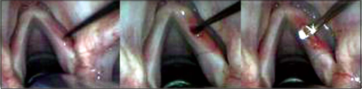
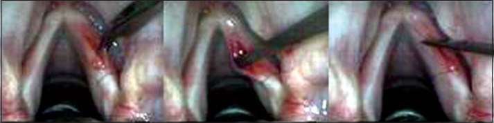
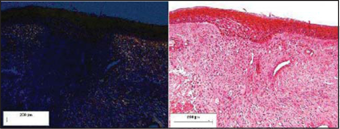
Many techniques were applied to treat patients with sulcus vocalis and scarred vocal folds. Their results were not good enough. In the Technique of Vocal Fold Pediculated Mucosa Flap, an anterior pediculated flap of vocal fold is positioned on the superficial layer of the lamina propria, below the free margin. Aim: To describe histological postoperative findings on the superficial layer of lamina propria during the application of the technique Vocal Fold Pediculated Mucosa Flap. The following parameters were compared between tested and control groups: total, type I and type III collagen and number of cellular nucleus. Study design: experimental. Material and Method: Fifteen dogs were used. One vocal fold was submitted to the intervention and the other was left as control. Each group of three dogs was sacrificed on 10, 30, 90, 180 and 360 days after the experimental surgery. Hematoxylin and eosin (H.E.) and Syrius Red were the staining techniques used. Results: Type I and total collagen suggested increased results in the tested group on postoperative days 90 and 180, nevertheless there was statistical significance only on postoperative day 180 (p<0.05). Type III collagen group area was less significant than the control group on postoperative day 180 (p<0.05). The number of cellular nucleus was increased on the 10th postoperative day, but decreased after the 30th day. Discussion: The findings about total and type I collagen and the amount of cellular nucleus on the superficial layer of lamina propria were similar to laryngeal postoperative studies in dogs. More complex studies would contribute with new data about the present subject.
Figures


Similar articles
-
Comparative histology and vibration of the vocal folds: implications for experimental studies in microlaryngeal surgery.Laryngoscope. 2000 May;110(5 Pt 1):814-24. doi: 10.1097/00005537-200005000-00011. Laryngoscope. 2000. PMID: 10807360
-
Histologic comparison of vocal fold microflap healing with sutures and glue.Laryngoscope. 2013 Jul;123(7):1709-16. doi: 10.1002/lary.23914. Epub 2013 Apr 26. Laryngoscope. 2013. PMID: 23625704
-
Classification for animal vocal fold surgery: resection margins impact histological outcomes of vocal fold injury.Laryngoscope. 2014 Nov;124(11):E437-44. doi: 10.1002/lary.24799. Epub 2014 Jun 26. Laryngoscope. 2014. PMID: 24965969 Free PMC article.
-
Vocal outcomes following subepithelial infiltration technique in microflap surgery: a review of 30 cases.J Laryngol Otol. 2007 Aug;121(8):768-71. doi: 10.1017/S002221510700744X. Epub 2007 Apr 20. J Laryngol Otol. 2007. PMID: 17445356 Review.
-
Implantation of atelocollagen sheet for vocal fold scar.Curr Opin Otolaryngol Head Neck Surg. 2010 Dec;18(6):507-11. doi: 10.1097/MOO.0b013e32833febdc. Curr Opin Otolaryngol Head Neck Surg. 2010. PMID: 20856118 Free PMC article. Review.
References
-
- Ishiki N, Tsuji DH, Sennes U. Tireoplastia. 1. Bios Comunicação e Editora Ltda.; São Paulo: 1999. pp. 1–49.
-
- Ford CN. Advances and Refinements in Phonosurgery. Laryngoscope. 1999;109:1891–1900. - PubMed
-
- Fisher KV, Telser A, Phillips JE, Yeates DB. Regulation of vocal fold transepithelial water fluxes. J Appl Physiol. 2001;91:1401–1411. - PubMed
-
- Junqueira LCU, Montes GS, Martins JEC. Dermal Collagen Distribution. A Histochemical and Ultrastructural Study. Histochemistry. 1983;79:397–403. - PubMed
-
- Benninger MS, Alessi D, Archer S, Bastian R, Ford C, Koufman J, Sataloff RT, Spiegel JR, Woo P. Vocal fold scarring: current concepts and management. Otolaryngology- Head and Neck Surgery. 1996;115(5):474–482. - PubMed
Publication types
MeSH terms
Substances
LinkOut - more resources
Full Text Sources
