. 2006 May 1;395(3):587-98.
doi: 10.1042/BJ20051965.

Reinvestigation of the dysbindin subunit of BLOC-1 (biogenesis of lysosome-related organelles complex-1) as a dystrobrevin-binding protein

Affiliations

Reinvestigation of the dysbindin subunit of BLOC-1 (biogenesis of lysosome-related organelles complex-1) as a dystrobrevin-binding protein

Ramin Nazarian et al. Biochem J. .

Abstract

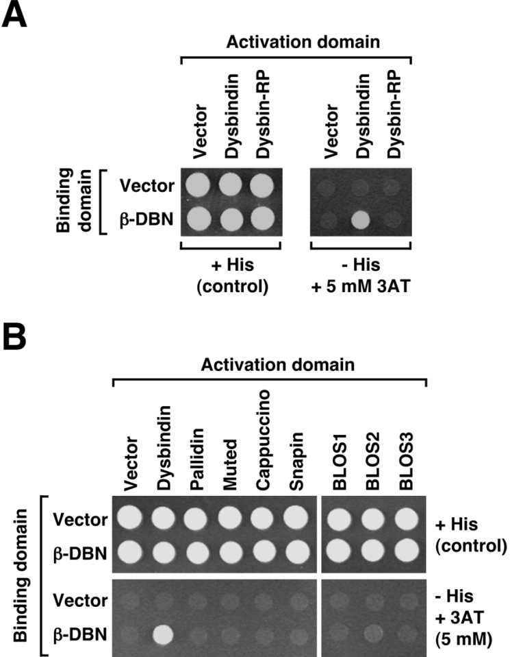
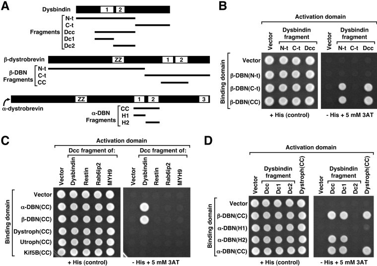
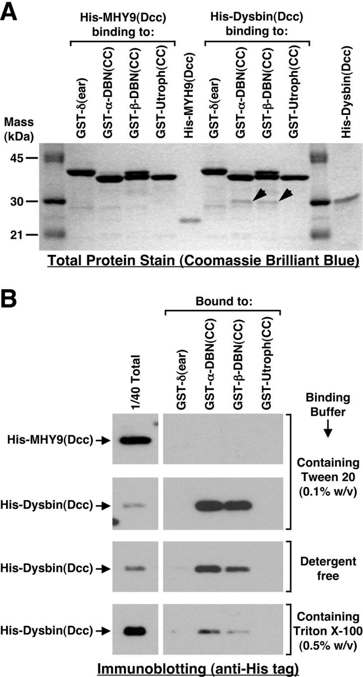
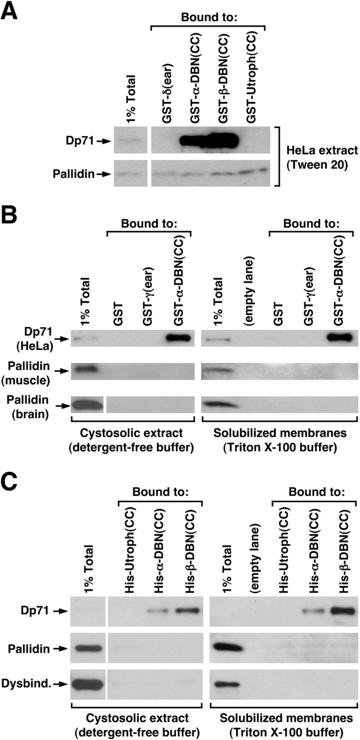
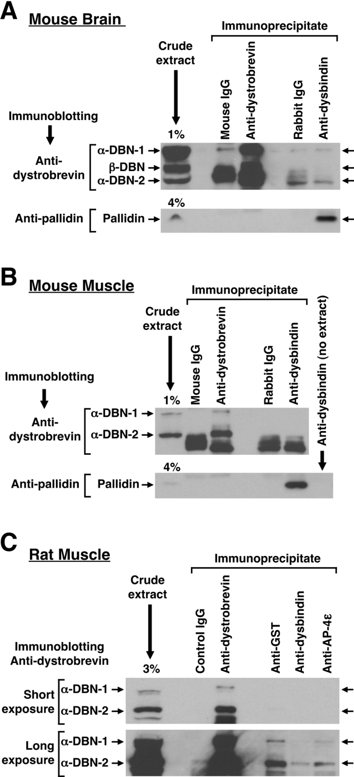
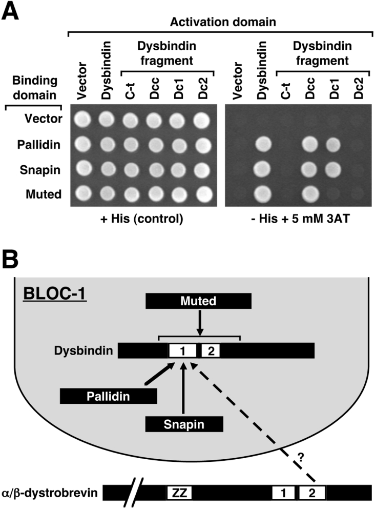
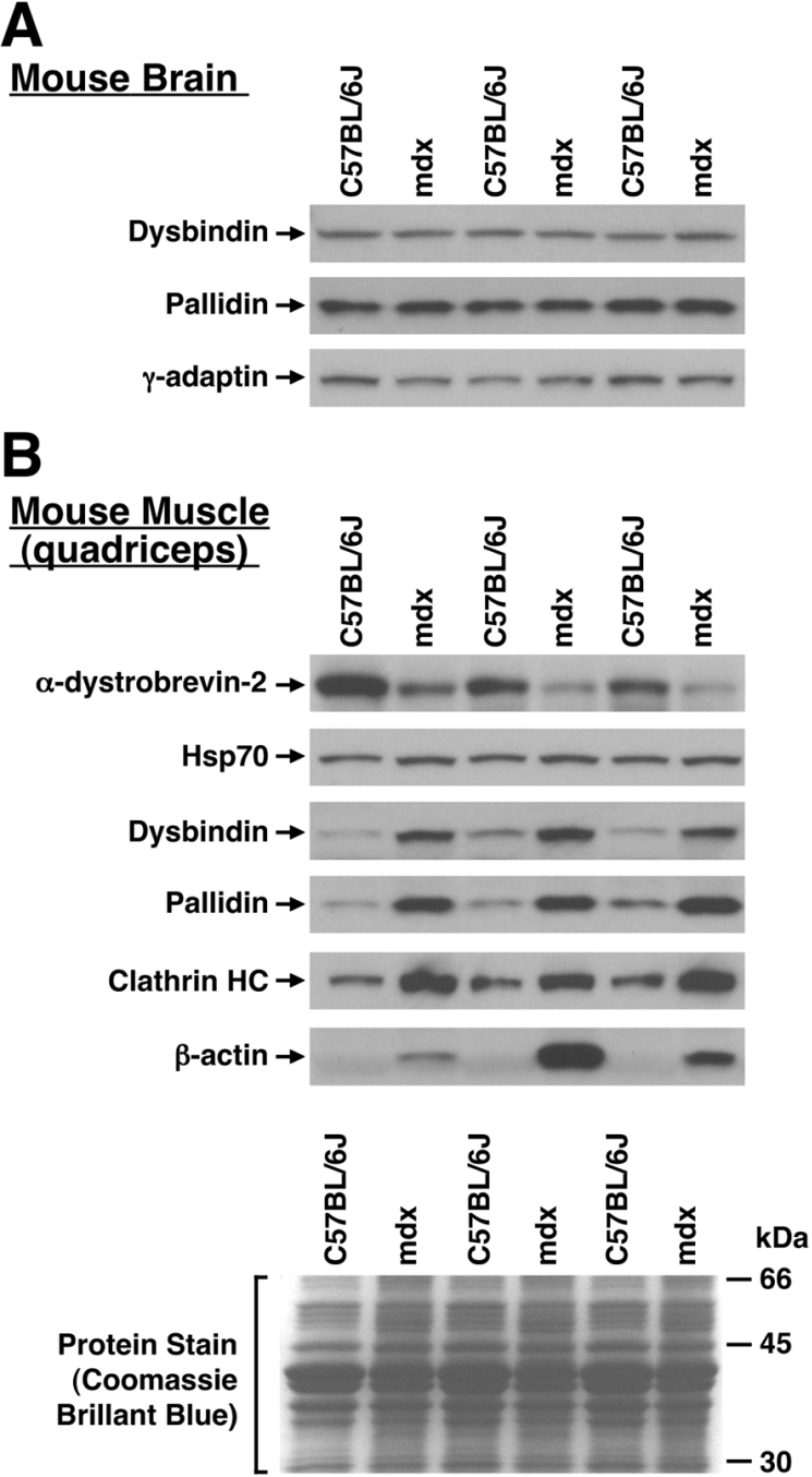
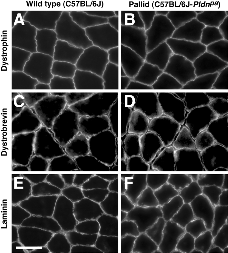
Dysbindin was identified as a dystrobrevin-binding protein potentially involved in the pathogenesis of muscular dystrophy. Subsequently, genetic studies have implicated variants of the human dysbindin-encoding gene, DTNBP1, in the pathogeneses of Hermansky-Pudlak syndrome and schizophrenia. The protein is a stable component of a multisubunit complex termed BLOC-1 (biogenesis of lysosome-related organelles complex-1). In the present study, the significance of the dystrobrevin-dysbindin interaction for BLOC-1 function was examined. Yeast two-hybrid analyses, and binding assays using recombinant proteins, demonstrated direct interaction involving coiled-coil-forming regions in both dysbindin and the dystrobrevins. However, recombinant proteins bearing the coiled-coil-forming regions of the dystrobrevins failed to bind endogenous BLOC-1 from HeLa cells or mouse brain or muscle, under conditions in which they bound the Dp71 isoform of dystrophin. Immunoprecipitation of endogenous dysbindin from brain or muscle resulted in robust co-immunoprecipitation of the pallidin subunit of BLOC-1 but no specific co-immunoprecipitation of dystrobrevin isoforms. Within BLOC-1, dysbindin is engaged in interactions with three other subunits, named pallidin, snapin and muted. We herein provide evidence that the same 69-residue region of dysbindin that is sufficient for dystrobrevin binding in vitro also contains the binding sites for pallidin and snapin, and at least part of the muted-binding interface. Functional, histological and immunohistochemical analyses failed to detect any sign of muscle pathology in BLOC-1-deficient, homozygous pallid mice. Taken together, these results suggest that dysbindin assembled into BLOC-1 is not a physiological binding partner of the dystrobrevins, likely due to engagement of its dystrobrevin-binding region in interactions with other subunits.


Figures

Figure 1
Figure 1. Interaction between human dysbindin and β-dystrobrevin in the context of the yeast two-hybrid system
(A) Analysis of interaction between β-dystrobrevin and dysbindin or dysbindin-related protein (Dysbin-RP). (B) Analysis of interaction between β-dystrobrevin (β-DBN) and the eight known subunits of BLOC-1. (A, B) Yeast cells were co-transformed with expression plasmids encoding Gal4 DNA-binding and activation domains alone (Vector) or fused in-frame to the full-length ORFs of the indicated human proteins. Double transformants were grown on minimal medium lacking leucine and tryptophan and containing histidine, suspended in distilled water at equal cell density, and then spotted on the same medium (+His) or on selective medium lacking histidine (−His) and containing 5 mM 3AT. Colony growth was examined after 3 days of culture at 30 °C.
Figure 2
Figure 2. Mapping of regions involved in the interaction between dysbindin and the dystrobrevins using the yeast two-hybrid system
(A) Schematic representation of the predicted domain organizations of dysbindin and the α- and β-dystrobrevins (DBNs), as well as of fragments used for mapping experiments. Coiled-coil-forming domains in each protein are denoted with numbers on a white background; notice that there is no significant identity between the coiled-coil-forming regions of dysbindin and those of the dystrobrevins. Both dystrobrevins also contain a ZZ-type zinc finger. (BD) Yeast cells were co-transformed with expression plasmids encoding Gal4 DNA-binding and activation domains alone (vector) or fused in-frame to the indicated human protein segments. The Dystroph(CC), Utroph(CC) and Kif5B(CC) constructs contain segments of dystrophin, utrophin and kinesin heavy chain Kif5B respectively, which display identity to the CC region of the dystrobrevins. Likewise, the ‘Dcc fragments’ of restin, Rab6ip2 and MYH9 are related to the Dcc region of dysbindin. Double transformants were spotted on minimal medium lacking leucine and tryptophan and containing histidine (+His) or lacking histidine and containing 5 mM 3AT (– His+5 mM 3AT). Colony growth was examined by visual inspection after 3 days of culture at 30 °C.
Figure 3
Figure 3. Interaction between recombinant proteins containing the coiled-coil-forming domains of dysbindin and the dystrobrevins
Purified GST-fusion proteins containing the coiled-coil-forming segments (CC) of human α-dystrobrevin (α-DBN), β-dystrobrevin (β-DBN) and utrophin (Utroph), as well as the irrelevant GST-δ(ear) protein, were immobilized on glutathione–Sepharose beads and incubated with purified polyhistidine-tagged-fusion proteins containing the dysbindin coiled-coil-forming region [His–Dysbin(Dcc)] or the corresponding region in MYH9 [His–MYH9(Dcc)]. Following incubation, the beads were isolated by centrifugation, washed extensively, and heated in the presence of SDS/PAGE sample buffer. Samples were analysed by SDS/PAGE followed by Coomassie Brilliant Blue staining (A) or immunoblotting using a monoclonal antibody to the polyhistidine tag (B). Arrowheads in (A) denote the presence of His–Dysbin(Dcc) protein associated with GST–α-DBN(CC) and GST–β-DBN(CC). In (B), each pair of ‘1/40 total’ and ‘Bound’ digital images is derived from portions of a single immunoblot that was exposed to a film, scanned and processed under identical conditions. Notice that the His–Dysbin(Dcc) protein specifically bound to both GST–α-DBN(CC) and GST–β-DBN(CC), although the relative amounts of bound His–Dysbin(Dcc) varied depending on the composition of the binding buffer. Further details on the buffer compositions are provided in the main text.
Figure 4
Figure 4. Affinity-pulldown binding experiments using cell-free extracts
(A, B) The indicated GST-fusion proteins were immobilized on glutathione–Sepharose beads and subsequently incubated with cell-free extracts freshly prepared from HeLa cells, mouse whole brain, or mouse quadriceps muscle, using the indicated buffers (see the text for buffer compositions and further experimental details). Following the incubation period, the beads were isolated by brief centrifugation, washed with ice-cold buffer, and then heated in the presence of SDS/PAGE sample buffer to elute the GST-fusion proteins and any associated proteins. Eluted protein samples were analysed by immunoblotting using monoclonal antibodies to dystrophin (to detect the Dp71 isoform as a positive control) or to the pallidin subunit of BLOC-1. (C) Polyhistidine-fusion proteins containing the S-tag sequence and the coiled-coil-forming domains (CC) of human α-dystrobrevin (α-DBN), β-dystrobrevin (β-DBN) and utrophin (Utroph) were immobilized on Protein S–agarose beads and subsequently incubated with freshly prepared cytosolic and solubilized membrane protein extracts from HeLa cells. Following incubation, the beads were recovered by centrifugation and washed. Bound proteins were eluted using SDS/PAGE sample buffer, and then analysed by immunoblotting using monoclonal antibodies to dystrophin/Dp71 and pallidin or a polyclonal antibody to dysbindin. Each image of the control lane represents 1% of the total extract and is from the same immunoblot exposed to a film, scanned and processed under identical conditions as the images corresponding to ‘Bound’ material. Notice in (C) that the relative amounts of Dp71 bound to His–α-DBN(CC) and His–β-DBN(CC) were such that the Dp71 protein band was only visible in the ‘1% Total’ control lane after a significantly longer exposure (not shown).
Figure 5
Figure 5. Co-immunoprecipitation experiments
Detergent extracts prepared from mouse brain (A), mouse quadriceps muscle (B) or rat quadriceps (C) were subjected to immunoprecipitation using a mouse monoclonal antibody against α- and β-dystrobrevins, a rabbit polyclonal antibody to dysbindin, and comparable amounts of the indicated control antibodies. The washed immunoprecipitates were analysed by immunoblotting using monoclonal antibodies to dystrobrevin or pallidin. Small aliquots (1–4% as indicated) of the crude extracts used for immunoprecipitation were analysed in parallel. Notice in (A, B) that significant amounts of pallidin were specifically recovered from the sample immunoprecipitated using anti-dysbindin, and that no dystrobrevin was found in the anti-dysbindin immunoprecipitate in amounts higher than in control immunoprecipitates. Similarly, only minute amounts of α-dystrobrevin isoforms were detected in the anti-dysbindin immunoprecipitate obtained from rat quadriceps extracts, and these amounts were not higher than those of dystrobrevins non-specifically associated with immunoprecipitates obtained using irrelevant rabbit polyclonal antibodies (C).
Figure 6
Figure 6. Mapping of dysbindin regions involved in interaction with other BLOC-1 subunits using the yeast two-hybrid system
(A) Yeast cells were co-transformed with expression plasmids encoding Gal4 DNA-binding and activation domains alone (vector), fused in-frame to the full-length ORFs of human dysbindin, pallidin, snapin and muted, or fused in-frame to the indicated fragments of human dysbindin. Double transformants were grown on minimal medium lacking leucine and tryptophan and containing histidine (+His), suspended in distilled water at an equal cell density, and then spotted on the same medium or on selective medium lacking histidine (−His) and containing 5 mM 3AT. Colony growth was examined after 3 days of culture at 30 °C. (B) Schematic representation of how the results shown in Figures 2(D), 3, 4 and 5 can be interpreted in light of the results shown in (A). For the sake of simplicity, only the relevant BLOC-1 subunits are shown, and the coiled-coil-forming regions of muted, pallidin and snapin are not depicted.
Figure 7
Figure 7. Immunoblot analysis of the relative contents of selected proteins in whole-tissue extracts prepared from wild-type and dystrophin-deficient mice
Extracts were prepared from brain (A) or quadriceps muscle (B) freshly dissected from 2-month-old male mice of wild-type (C57BL/6J) or dystrophin-deficient (mdx) strains (3 mice per strain). The extracts (60 μg of total protein) were analysed by SDS/PAGE, followed by immunoblotting using specific antibodies against the indicated proteins (clathrin HC, clathrin heavy chain). The lower panel in (B) shows a portion of a duplicate SDS/polyacrylamide gel that was stained using Coomassie Brilliant Blue.
Figure 8
Figure 8. Immunohistochemical analyses of muscle sections from wild-type and BLOC-1-deficient (pallid) mice
Sections of quadriceps dissected from 4-month-old female mice of the indicated strains were stained using antibodies to dystrophin (A, B), dystrobrevin (C, D) and laminin (E, F) followed by appropriate secondary antibodies conjugated with Texas Red (A, B) or fluorescein (CF). Scale bar, 50 μm.

Similar articles

Cited by

References

    1. Blake D. J., Weir A., Newey S. E., Davies K. E. Function and genetics of dystrophin and dystrophin-related proteins in muscle. Physiol. Rev. 2002;82:291–329. - PubMed
    1. Albrecht D. E., Froehner S. C. Syntrophins and dystrobrevins: defining the dystrophin scaffold at synapses. Neurosignals. 2002;11:123–129. - PubMed
    1. Durbeej M., Campbell K. P. Muscular dystrophies involving the dystrophin–glycoprotein complex: an overview of current mouse models. Curr. Opin. Genet. Dev. 2002;12:349–361. - PubMed
    1. Blake D. J. Dystrobrevin dynamics in muscle-cell signaling: a possible target for therapeutic intervention in Duchenne muscular dystrophy? Neuromuscul. Disord. 2002;12:S110–S117. - PubMed
    1. Grady R. M., Grange R. W., Lau K. S., Maimone M. M., Nichol M. C., Stull J. T., Sanes J. R. Role for α-dystrobrevin in the pathogenesis of dystrophin-dependent muscular dystrophies. Nat. Cell Biol. 1999;1:215–220. - PubMed

Publication types

MeSH terms

Associated data