. 2006 Jan 30;172(3):423-32.
doi: 10.1083/jcb.200505175.

P21-activated kinase 1: convergence point in PDGF- and LPA-stimulated collagen matrix contraction by human fibroblasts

Affiliations

P21-activated kinase 1: convergence point in PDGF- and LPA-stimulated collagen matrix contraction by human fibroblasts

Sangmyung Rhee et al. J Cell Biol. .

Abstract

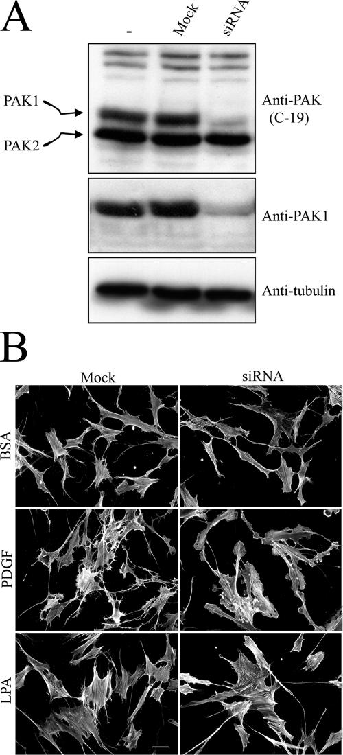
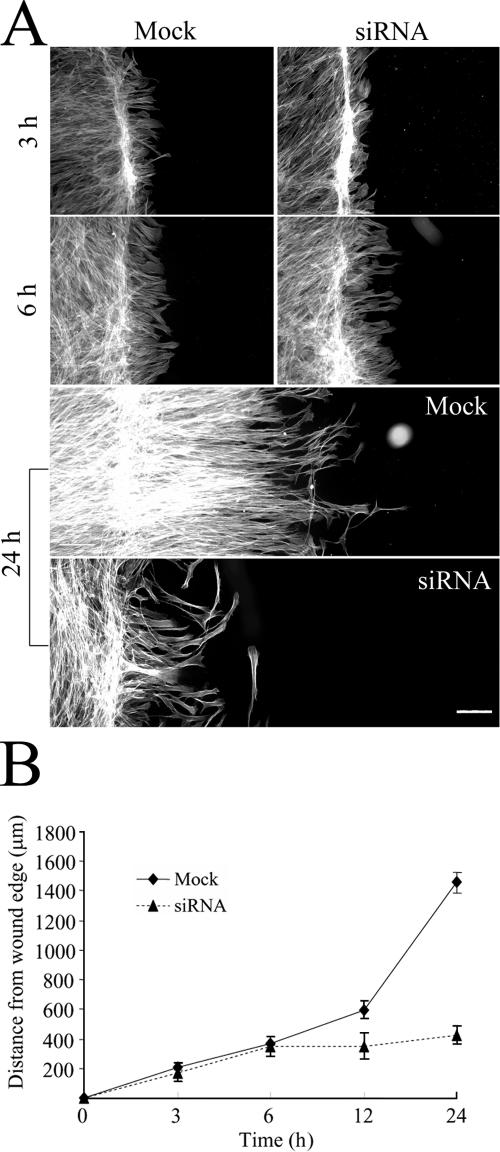
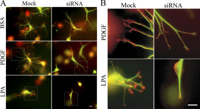
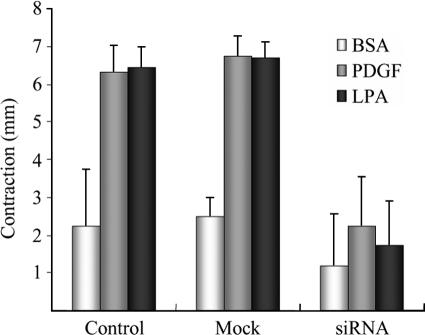
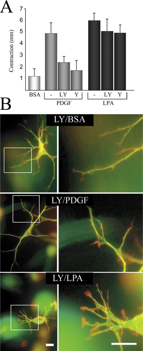
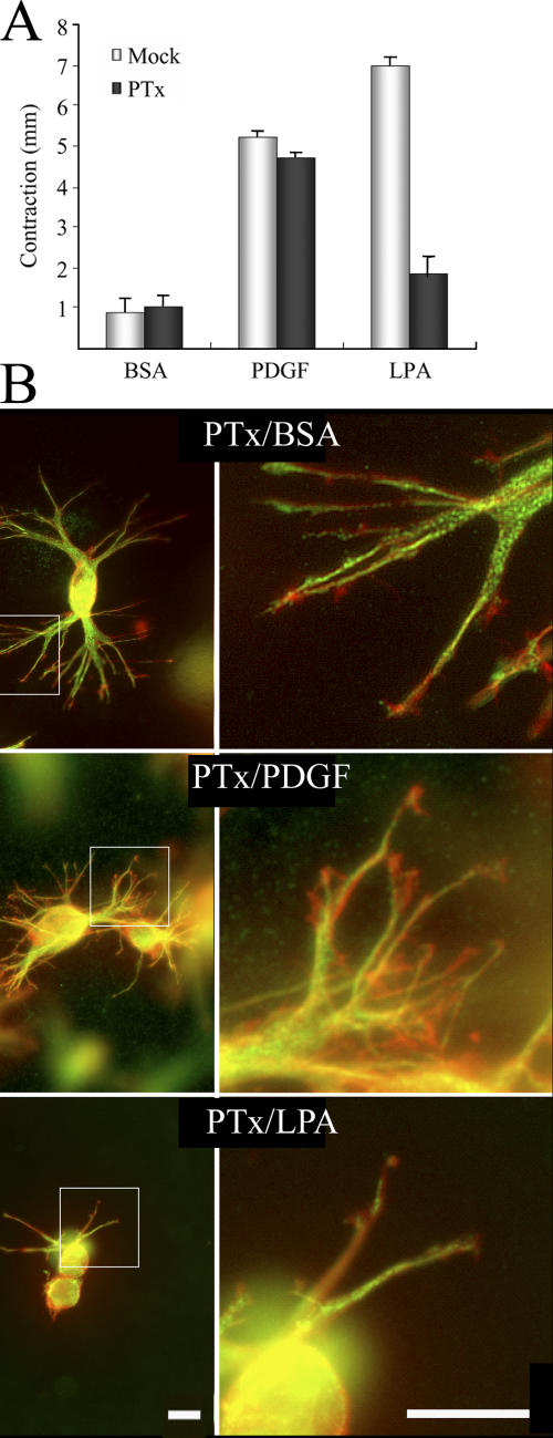
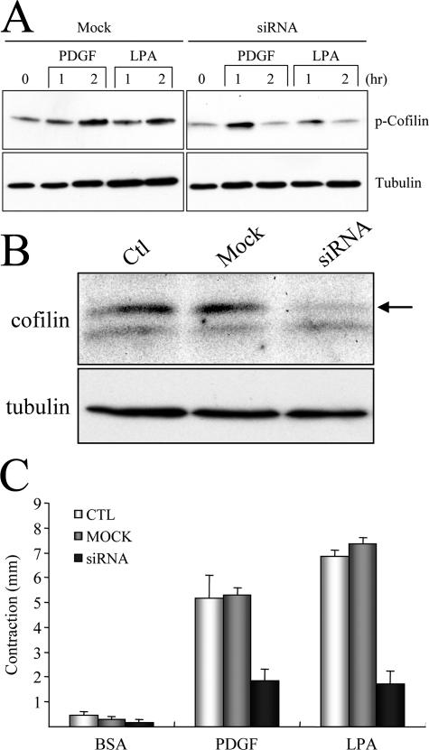
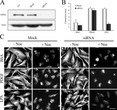
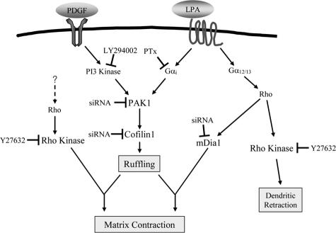
Fibroblast three-dimensional collagen matrix culture provides a tissue-like model that can be used to analyze cell form and function. The physiological agonists platelet-derived growth factor (PDGF) and lysophosphatidic acid (LPA) both stimulate human fibroblasts to contract floating collagen matrices. In this study, we show that the PDGF and LPA signaling pathways required for matrix contraction converge on p21-activated kinase 1 (PAK1) and its downstream effector cofilin1 and that contraction depends on cellular ruffling activity, rather than on the protrusion and retraction of cellular dendritic extensions. We also show that, depending on the agonist, different Rho effectors cooperate with PAK1 to regulate matrix contraction, Rho kinase in the case of PDGF and mDia1 in the case of LPA. These findings establish a unified framework for understanding the cell signaling pathways involved in fibroblast contraction of floating collagen matrices.


Figures

Figure 1.
Figure 1.
PAK1 silencing in human fibroblasts and cell morphology. (A) Cells were transfected for 12 h with 700 nM siRNA or sense RNA only (Mock) and cultured for an additional 24 h in growth medium without siRNA. Extracts were prepared and subjected to SDS-PAGE and immunoblotted to analyze levels of PAK1, PAK2, and tubulin. (B) PAK1-silenced and mock-transfected cells were harvested and incubated for 1 h on collagencoated glass coverslips in DME containing 5 mg/ml BSA and 10 μM LPA or 50 ng/ml PDGF as indicated. At the end of the incubation, samples were fixed and stained for actin. Bar, 50 μm.
Figure 2.
Figure 2.
Silencing PAK1 inhibits cell migration. (A) PAK1-silenced and mock-transfected cells were harvested and cultured overnight on collagen-coated coverslips. After scrape wounding, the cultures were incubated in DME containing 5 mg/ml BSA and 50 ng/ml PDGF. At the end of the incubations, samples were fixed and stained for actin. (B) Migration was quantified by determining the average distance of cell migration from the wound edge based on measurement of 10 separate microscopic fields and 5 cells within each field. Bar, 150 μm.
Figure 3.
Figure 3.
Dendritic extensions and ruffling by PAK1-silenced human fibroblasts incubated for 1 h in collagen matrices. (A) PAK1-silenced and mock-transfected cells were harvested and used to prepare floating collagen matrices. Samples were incubated for 1 h in DME containing 5 mg/ml BSA and 50 ng/ml PDGF or 10 μM LPA, as indicated. At the end of the incubations, samples were fixed and stained with anti-tubulin antibody for microtubules and rhodamine-phalloidin for actin. (B) Enlarged view of dendritic extension tips in the boxed areas. Green, microtubules; red, actin. Bar, 20 μm.
Figure 4.
Figure 4.
Dendritic extensions and ruffling by PAK1-silenced fibroblasts incubated for 4 h in collagen matrices. (A) PAK1-silenced and mock-transfected cells were harvested and used to prepare floating collagen matrices. Samples were incubated for 4 h in DME containing 5 mg/ml BSA and 50 ng/ml PDGF or 10 μM LPA, as indicated. At the end of the incubations, samples were fixed and stained with anti-tubulin antibody for microtubules and rhodamine-phalloidin for actin. (B) Enlarged view of dendritic extension tips in the boxed areas. Green, microtubules; red, actin. Bar, 20 μm.
Figure 5.
Figure 5.
Inhibition of collagen matrix contraction in PAK1-silenced cells. Nontransfected (Control), PAK1-silenced, and mock-transfected cells were harvested and used to prepare floating collagen matrices. Samples were incubated for 4 h in DME with 5 mg/ml BSA and 50 ng/ml PDGF or 10 μM LPA added as shown. At the end of the incubations samples were fixed and the extent of matrix contraction was measured as the decrease in matrix diameter. Data shown are arithmetic mean ± SD for three separate experiments.
Figure 6.
Figure 6.
PI3 kinase and Rho kinase are required for PDGF-stimulated, but not LPA-stimulated, collagen matrix contraction. (A) Fibroblasts were harvested and used to prepare floating collagen matrices. Samples were incubated for 4 h in DME with 5 mg/ml BSA and 50 ng/ml PDGF, 10 μM LPA, 20 μM LY294002 (LY; PI3 kinase inhibitor), and 10 μM Y27634 (Y; Rho kinase inhibitor) added where indicated. At the end the incubations, samples were fixed and the extent of matrix contraction was measured as the decrease in matrix diameter. Data shown are arithmetic means ± SD for three separate experiments. (B) Selected samples from A were fixed and stained with anti-tubulin antibody for microtubules (green) and rhodaminephalloidin for actin (red). Enlarged view of dendritic extension tips in the boxed areas. Bar, 20 μm.
Figure 7.
Figure 7.
Pertussis toxin inhibits LPA-stimulated, but not PDGF-stimulated, collagen matrix contraction. (A) Fibroblasts in monolayer culture were incubated overnight with 25 ng/ml pertussis toxin. Subsequently, the cells were harvested and used to prepare floating collagen matrices. Samples were incubated for 4 h in DME with 5 mg/ml BSA and 50 ng/ml PDGF or 10 μM LPA added where indicated. At the end the incubations, samples were fixed and the extent of matrix contraction was measured as the decrease in matrix diameter. Data shown are arithmetic means ± SD for three separate experiments. (B) Selected samples from A were fixed and stained with anti-tubulin antibody for microtubules (green) and rhodamine-phalloidin for actin (red). (right) Enlarged view of dendritic extension tips in the boxed areas. Bars, 20 μm.
Figure 8.
Figure 8.
Cofilin1 is downstream of PAK1 in LPA and PDGF regulation of collagen matrix contraction. (A) PAK1-silenced and mock-transfected cells were harvested and used to prepare floating collagen matrices. Samples were incubated for the times shown in DME containing 5 mg/ml BSA and 50 ng/ml PDGF or 10 μM LPA, as indicated. At the end of the incubations, extracts of the samples were prepared and subjected to immunoblotting with antibodies directed against phospho-cofilin1 and tubulin. (B) Cells were transfected for 36 h with 500 nM of cofilin1 siRNA or sense RNA only (Mock) and then cultured an additional 24 h in growth medium without siRNA. Extracts were prepared and subjected to SDS-PAGE and immunoblotted to analyze levels of cofilin1 (arrow) and tubulin. (C) Nontransfected (CTL), cofilin1-silenced, and mock-transfected cells were harvested and used to prepare floating collagen matrices. Samples were incubated for 4 h in DME with 5 mg/ml BSA and 50 ng/ml PDGF or 10 μM LPA added as shown. At the end of the incuba-tions, samples were fixed and the extent of matrix contraction was meas-ured as the decrease in matrix diameter. Data shown are arithmetic means ± SD for three separate experiments.
Figure 9.
Figure 9.
mDia1 cooperates with PAK1 in LPA regulation of collagen matrix contraction silencing in human fibroblasts. (A) Cells were transfected for 12 h with 700 nM siRNA or sense RNA only (Mock) and cultured an additional 24 h in growth medium without siRNA. Extracts were prepared and subjected to SDS-PAGE and immunoblotted to analyze levels of mDia1 and actin. (B) mDia1-silenced and mock-transfected cells were harvested and used to prepare floating collagen matrices. Samples were incubated for 4 h in DME with 5 mg/ml BSA and 50 ng/ml PDGF or 10 μM LPA added as shown. At the end of the incubations, samples were fixed, and the extent of matrix contraction was measured as the decrease in matrix diameter. Data shown are arithmetic means ± SD for three separate experiments. (C) At the end of the transfection period, mock- and siRNA-transfected cells were incubated in serum-free medium for 36 h, followed by 4 h in DME with 5 mg/ml BSA and 50 ng/ml PDGF or 10 μM LPA added as shown. Stable microtubules were detected as previously described (Gundersen et al., 1994; Cook et al., 1998). Subsequently, the indicated samples were treated with 2 μM nocodazole for 2 h . After two rinses with microtubule-stabilizing buffer (MSB; 85 mM Pipes, pH 6.9, 1 mM EGTA, 1 mM MgCl2, 2 M glycerol, 1 μg/ml leupeptin, 1 μg/ml pepstatin A, and 1 mM 4-(2-aminoethyl)-benzenesulfonyl fluoride), samples were treated with 1 ml of MSB containing 200 μg/ml saponin for 5 min at 37°C to extract tubulin monomer, rinsed with MSB, and fixed with methanol (−20°C) for 10 min, and then stained with anti-tubulin antibody. Bar, 50 μm.
Figure 10.
Figure 10.
Signaling pathways in floating collagen matrix contraction. Model showing convergence of PDGF and LPA signaling on PAK1 and cofilin1, cell ruffling, and collagen matrix contraction. Rho kinase cooperates with PAK1 for PDGF-stimulated contraction, whereas mDia1 cooperates with PAK1 for LPA-stimulated contraction. Rho kinase also is required for LPA-stimulated retraction of dendritic extensions.

Similar articles

Cited by

References

    1. Abe, M., C.H. Ho, K.E. Kamm, and F. Grinnell. 2003. Different molecular motors mediate platelet-derived growth factor and lysophosphatidic acid-stimulated floating collagen matrix contraction. J. Biol. Chem. 278:47707–47712. - PubMed
    1. Ahlen, K., A. Berg, F. Stiger, A. Tengholm, A. Siegbahn, E. Gylfe, R.K. Reed, and K. Rubin. 1998. Cell interactions with collagen matrices in vivo and in vitro depend on phosphatidylinositol 3-kinase and free cytoplasmic calcium. Cell Adhes. Commun. 5:461–473. - PubMed
    1. Anliker, B., and J. Chun. 2004. Lysophospholipid G protein-coupled receptors. J. Biol. Chem. 279:20555–20558. - PubMed
    1. Arber, S., F.A. Barbayannis, H. Hanser, C. Schneider, C.A. Stanyon, O. Bernard, and P. Caroni. 1998. Regulation of actin dynamics through phosphorylation of cofilin by LIM-kinase. Nature. 393:805–809. - PubMed
    1. Bamburg, J.R. 1999. Proteins of the ADF/cofilin family: essential regulators of actin dynamics. Annu. Rev. Cell Dev. Biol. 15:185–230. - PubMed

Publication types

MeSH terms

Substances