. 2006 Jan 30;172(3):453-67.
doi: 10.1083/jcb.200506136.

Localized recruitment and activation of RhoA underlies dendritic spine morphology in a glutamate receptor-dependent manner

Affiliations

Localized recruitment and activation of RhoA underlies dendritic spine morphology in a glutamate receptor-dependent manner

Vanessa Schubert et al. J Cell Biol. .

Abstract

Actin is the major cytoskeletal source of dendritic spines, which are highly specialized protuberances on the neuronal surface where excitatory synaptic transmission occurs (Harris, K.M., and S.B. Kater. 1994. Annu. Rev. Neurosci. 17:341-371; Yuste, R., and D.W. Tank. 1996. Neuron. 16:701-716). Stimulation of excitatory synapses induces changes in spine shape via localized rearrangements of the actin cytoskeleton (Matus, A. 2000. Science. 290:754-758; Nagerl, U.V., N. Eberhorn, S.B. Cambridge, and T. Bonhoeffer. 2004. Neuron. 44:759-767). However, what remains elusive are the precise molecular mechanisms by which different neurotransmitter receptors forward information to the underlying actin cytoskeleton. We show that in cultured hippocampal neurons as well as in whole brain synaptosomal fractions, RhoA associates with glutamate receptors (GluRs) at the spine plasma membrane. Activation of ionotropic GluRs leads to the detachment of RhoA from these receptors and its recruitment to metabotropic GluRs. Concomitantly, this triggers a local reduction of RhoA activity, which, in turn, inactivates downstream kinase RhoA-specific kinase, resulting in restricted actin instability and dendritic spine collapse. These data provide a direct mechanistic link between neurotransmitter receptor activity and the changes in spine shape that are thought to play a crucial role in synaptic strength.


Figures
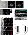
Figure 1.
Figure 1.
High concentrations of KCl lead to local depolymerization of F-actin and dendritic spine retraction. (A) Dendritic spines were visualized with phalloidin (F-actin; red in merge) and presynaptic terminals labeled with synaptotagmin (ST; blue in merge). Close apposition between postsynaptic F-actin accumulations and presynaptic terminals is observed (panels 1 and 2). (B) DiI (green in merge)-labeled neurons were incubated with 5 or 55 mM KCl before fixation and labeled with phalloidin (blue in merge). The F-actin accumulations on the dendritic surface coincide with the membranous protrusions detected with DiI (5 mM KCl; arrowheads). Upon 55 mM KCl treatment (55 mM KCl), DiI and F-actin labeling reveal the absence of actin-rich membranous protrusions and a rather high accumulation of F-actin in the dendritic shaft compared with controls (5 mM KCl). (C) Immunofluorescence analysis of hippocampal neurons treated with 5 or 55mM KCl before fixation. Untreated and control buffer-treated (5 mM KCl) cells show similar numbers of dendritic spines, whereas 55 mM KCl-treated neurons exhibit an 85% reduction of dendritic spine number as judged by F-actin–rich protrusions at the surface (graph a). Replacing neurons into growth medium (55 mM KCl + wash) allowed a nearly complete recovery of spine number (graph b). Error bars represent SD. ***, P < 0.001; one-way ANOVA followed by Tukey's post-hoc test. Bars, 5 μm.
Figure 2.
Figure 2.
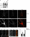
RhoA and F-actin behave similarly upon 55 mM KCl treatment. (A) RhoA is detectable in cytosol, synaptosomes, and in postsynaptic density fraction (PSD I). The enrichment of PSD 95 in respect to presynaptic protein synaptophysin demonstrates the pureness of the postsynaptic fraction. (B) Confocal slice of immunofluorescence analysis of RhoA localization to the dendritic spine. The cells were detergent extracted to visualize only membrane-bound and, thus, active RhoA. In untreated or control buffer–treated cells, RhoA localizes to the actin-rich domains of the cell (control). Enlargement of a dendritic segment revealed that apart from being present throughout the dendritic shaft, RhoA colocalizes with dendritic spine actin (panel 1, arrows). Note the 55 mM KCl-induced loss of dendritic spine actin and accumulation of F-actin cables in the shaft (55 mM KCl). In these cells, compared with controls, RhoA and F-actin signal intensity are greatly increased (graph). Error bars represent SD. The enlarged dendritic segment illustrates the colocalization between RhoA and F-actin in the dendritic shaft (panel 2; arrows indicate regions of colocalization). RhoA is green and F-actin is red in merged images. *, P < 0.001; paired t test. Bars, 10 μm.
Figure 3.
Figure 3.
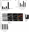
RhoA activity in synaptosomes is reduced upon 55 mM KCl treatment in a GluR-dependent manner. (A) Active RhoA (RhoA-GTP) was isolated from synaptosomes and whole cell extracts using a RhoA activation assay (AA). 55 mM KCl significantly decreased RhoA activity in synaptosomes compared with control buffer treatment (5 mM KCl). On the other hand, treatment of whole cells with 55 mM KCl largely increased RhoA activity in respect to controls. (B) Activation assay. Preincubation with MK-801 and CNQX prevented the 55 mM KCl-induced decrease of synaptosomal RhoA activity. (C) Dendritic segments showing GluR1 localization in mature neurons. In control cells, GluR1 labeling (green in merge) was punctate, and the protein colocalized with F-actin (red in merge)–rich dendritic spines (5 mM KCl, arrowheads). Upon 55 mM KCl treatment, F-actin cables formed in the shaft, and spine number was reduced (55 mM KCl, graph). Preincubation with CNQX or MK-801 prevented the spine loss and the formation of actin cables (graph). (D) Graph showing total spine number versus the number of spines containing GluR1 labeling. In cells treated with 5 and 55 KCl or CNQX + 55 mM KCl, the percentages were comparable (96–100%), whereas MK-801 + 55 mM KCl led to a 30% reduction of GluR1-containing spines (white arrowheads in C indicate GluR1-positive spines). Error bars represent SD. *, P < 0.001; paired t test. ***, P < 0.001; one-way ANOVA followed by Tukey's multiple comparison test. Bars, 5 μm.
Figure 4.
Figure 4.
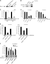
Activity of iGluRs determines the amount of receptor interaction with RhoA. (A) RhoA was coprecipitated with NMDAR2a and GluR1, respectively, from synaptosomal preparations. (B) Immunoprecipitation of RhoA from synaptosomes. The spine resident membrane proteins NMDA receptor 2a (NMDAR2a) and glutamate receptor 1 (GluR1) coprecipitate with RhoA. Incubation with GTPγS before the immunoprecipitation increased the interacttions to RhoA, whereas preincubation with GDP had the opposite effect. (C) RhoA activation assay. Reduced RhoA activity upon high potassium treatment (Fig. 3 A) diminishes coprecipitation of NMDAR2 and GluR1 with active RhoA (graph). *, P < 0.001; paired t test. (D) Less GluR1 and NMDAR2a coimmunoprecipitate with RhoA in synaptosomes after 55 mM KCl compared with control- (graph) or 5 mM KCl-treated samples. Similar effects are observed after direct stimulation with AMPA or NMDA, respectively. (E) Preincubation with specific antagonists for NMDA and GluRs MK-801 and CNQX, respectively, prevented the decrease of RhoA–GluR interaction induced by 55 mM KCl. (F) Biochemical analysis of the cross reactivity between GluR1 and NMDAR2a. Preincubation with NMDA antagonist MK-801 partially prevented the AMPA-induced reduction of GluR1–RhoA interaction (compare AMPA with MK-801 + AMPA). Furthermore, direct stimulation with NMDA decreased the amount of GluR1–RhoA interaction, and this could partially be blocked by CNQX (compare NMDA with CNQX + NMDA). On the other hand, stimulation with AMPA lowered RhoA–NMDAR2a interaction levels and, as such, could be prevented by preincubation with MK-801. Error bars represent SD. ***, P < 0.001; one-way ANOVA followed by Tukey's multiple comparison tests.
Figure 5.
Figure 5.
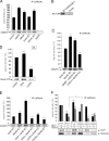
Association of RhoA with mGluR1 depends on AMPA receptor activation and is independent of RhoA activity levels. (A) mGluR1 coprecipitates with RhoA from rat synaptosomal preparations. Stimulation with AMPA, NMDA, or 55 mM KCl increased the interaction of mGluR1with RhoA. (B) Immunoprecipitation of mGluR1 from synaptosomes leads to coprecipitation of RhoA. (C) CNQX but not MK-801 prevents the 55 mM KCl-induced amplification of RhoA–mGluR1 interaction. (D) Activation assay performed on synaptosomes. Incubation with mGluR1 antagonist (AIDA) increases RhoA activity, whereas the agonist (DHPG) lowers activity levels of RhoA. (E) Effect of AIDA and DHPG on RhoA affinity to mGluR1. In respect to controls (5 mM KCl), incubation of synaptosomes with AIDA (AIDA + 5 mM KCl) and DHPG (DHPG + 5 mM KCl) increases the amount of receptor–RhoA interaction. Although the effect of AIDA appears very drastic, DHPG only slightly increases the RhoA–mGluR1 interaction. In both cases (AIDA and DHPG), subsequent treatment with 55 mM KCl leads to the opposite effect, inducing a drastic reduction of RhoA–mGluR1 interaction levels (AIDA + 55 mM KCl and DHPG + 55 mM KCl). (F) Effect of mGluR1 activity on AMPA and NMDAR interaction with RhoA. RhoA–GluR1 interaction was slightly (118%) increased by AIDA (AIDA + 5 mM KCl). DHPG did not have any significant effect (DHPG + 5 mM KCl). In both cases, stimulation with 55 mM KCl led to a detachment between RhoA and GluR1 (compare AIDA + 55 mM KCl and DHPG + 55 mM KCl with 55 mM KCl). AIDA slightly decreased the amount of NMDAR2a precipitating with RhoA under control conditions (compare AIDA + 5 mM KCl with 5 mM KCl). NMDAR2a resisted the 55-mM KCl stimulus in AIDA-treated samples, as NMDAR2a–RhoA interaction levels remained elevated (compare AIDA + 55 mM KCl and 55 mM KCl). Although simple DHPG treatment drastically reduced the amount of NMDAR2a–RhoA interaction (compare DHPG + 5 mM KCl and 5 mM KCl), subsequent incubation with 55 mM KCl led to interaction levels similar to controls (compare DHPG + 55 mM KCl and 55 mM KCl). Error bars represent SD. ***, P < 0.001; one way ANOVA followed by Tukey's multiple comparison tests. *, P < 0.001; paired t test.
Figure 6.
Figure 6.
RhoA-mediated dendritic spine F-actin stability is regulated in a ROCK–PIIa-dependent manner. (A) Analysis of subcellular fractionation of adult rat brain showed that ROCK and PIIa are detectable in cytosol, synaptosomes, and in PSD I fractions. (B) Representative confocal slice images of mature hippocampal neurons labeled for ROCK (a; green in merge) and profilinIIa (PIIa, b; green in merge). Both proteins are present throughout the cell, preferentially in F-actin–rich regions (red in merge). Enlargement of dendritic segments revealed that apart from being present throughout the dendritic shaft, ROCK and PIIa colocalize with dendritic spine actin. Boxed areas in whole cell images mark the localization of enlarged dendritic segments shown in the figure. Arrowheads indicate points of colocalization between F-actin and ROCK or PIIa, respectively. (C) ROCK and PIIa both coimmunoprecipitate with RhoA from synaptosomal fractions, and such interaction is favored by RhoA activation (GTPγS) and reduced if RhoA is inactivated (GDP). (D) 55 mM KCl largely inactivates RhoA compared with control buffer–treated samples (5 mM KCl; see Fig. 2 C). Concomitantly, the amount of coprecipitated ROCK and PIIa with active RhoA (RhoA-GTP) is drastically reduced, whereas the quantity of coprecipitated Diaphanous (Dia1) is not altered. Error bars represent SD. *, P < 0.001; paired t test. Bars, 5 μm.
Figure 7.
Figure 7.
Restrictive activation of caged ROCK inhibitor Y27632 induces localized spine retraction. Single images of in vitro time-lapse experiments performed on DiI-labeled neurons incubated with caged ROCK inhibitor (caged Y27632). (A) Images of a dendritic segment (dendrite 1) before (top) and after (1–8 min) uncaging of Y27632 by exposure to UV light. Dendritic spines retract upon activation of the compound (arrowheads). (B) A neighboring dendrite located outside the pinhole during uncaging of dendrite 1. The spines on this dendrite remain stable and do not retract (arrow-heads). Compare top image (before uncaging of dendrite 1) with bottom image (after uncaging and observation of dendrite 1). (C) Dendritic segment of a DiI-labeled neuron treated with the vehicle buffer used to dissolve the caged compound. UV stimulation did not induce changes in spines. Compare top image with the four bottom images (arrowheads). Bars, 5 μm.
Figure 8.
Figure 8.
Model describing the differential RhoA-related behavior of GluRs in response to excitatory stimuli. (A) Under steady-state conditions, active RhoA interacts with all three neurotransmitter receptor types at the level of the excitatory PSD, thus stabilizing spine actin filaments via its downstream effector complex ROCK–PIIa (here referred to as RhoA for simplification). Stimulation of neurotransmitter receptors at the excitatory PSD reduces the amount of total synaptic RhoA activity and leads to the redistribution of RhoA interactions away from iGluR toward mGluRs. This leads to a localized destabilization of dendritic spine actin, consequently allowing for actin-based plasticity and the instability of iGluRs at the PSD. This, in turn, would allow for the regulation of excitability of the synapse via either endocytosis and recycling of iGluRs, possibly induced by mGluR1 signaling, or the lateral movement of the receptors away from the PSD. Moreover, the stabilization of mGluR1 at the plasma membrane could guarantee signaling events, which are necessary for the cell to adjust to the incoming stimuli, such as the regulation of gene expression and modulation of cell survival pathways. Dashed boxes in both the control situation and excitatory stimulation of PSD panels represent localization of the excitatory synapse illustrated in A, B1, and B2. (B) Model illustrating how mGluR1 modulates NMDAR response to excitatory stimuli. (B1) Without active mGluR1 present, NMDARs become immune to incoming excitatory stimuli. Upon stimulation, high amounts of RhoA remain attached to the receptor, possibly leading to the retention of this receptor at the PSD. (B2) Activation of mGluR1 with DHPG has the opposite effect. The amount of RhoA–NMDAR interaction is greatly reduced and most likely leads to destabilization of NMDAR at the excitatory PSD, consequently reducing NMDA excitability.

Similar articles

Cited by

References

    1. Baskys, A., and M. Blaabjerg. 2005. Understanding regulation of nerve cell death by mGluRs as a method for development of successful neuroprotective strategies. J. Neurol. Sci. 229-230:201–209. - PubMed
    1. Blaabjerg, M., L. Fang, J. Zimmer, and A. Baskys. 2003. Neuroprotection against NMDA excitotoxicity by group I metabotropic glutamate receptors is associated with reduction of NMDA stimulated currents. Exp. Neurol. 183:573–580. - PubMed
    1. Bonhoeffer, T., and R. Yuste. 2002. Spine motility. Phenomenology, mechanisms, and function. Neuron. 35:1019–1027. - PubMed
    1. Bradke, F., and C.G. Dotti. 1997. Videomicroscopy of living microinjected hippocampal neurons in culture. In Microinjection and Transgenesis: Strategies and Protocols. A. Cid-Arregui and A. Garcia-Carrancà, editors. Springer-Verlag, Berlin, Germany. 81–94.
    1. Carlin, R.K., D.J. Grab, R.S. Cohen, and P. Siekevitz. 1980. Isolation and characterization of postsynaptic densities from various brain regions: enrichment of different types of postsynaptic densities. J. Cell Biol. 86:831–845. - PMC - PubMed

Publication types

MeSH terms

Substances