. 2006 Feb 7;103(6):1936-40.
doi: 10.1073/pnas.0510710103. Epub 2006 Jan 31.

Phospholipase D1 corrects impaired betaAPP trafficking and neurite outgrowth in familial Alzheimer's disease-linked presenilin-1 mutant neurons

Affiliations

Phospholipase D1 corrects impaired betaAPP trafficking and neurite outgrowth in familial Alzheimer's disease-linked presenilin-1 mutant neurons

Dongming Cai et al. Proc Natl Acad Sci U S A. .

Abstract

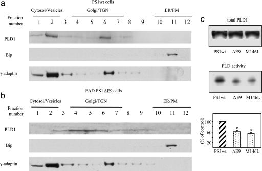
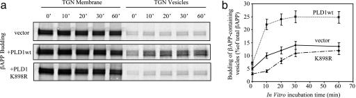
Presenilins (PS1/PS2) regulate proteolysis of beta-amyloid precursor protein (betaAPP) and affect its intracellular trafficking. Here, we demonstrate that a PS1-interacting protein, phospholipase D1 (PLD1), affects intracellular trafficking of betaAPP. Overexpression of PLD1 in PS1wt cells promotes generation of betaAPP-containing vesicles from the trans-Golgi network. Conversely, inhibition of PLD1 activity by 1-butanol decreases betaAPP trafficking in both wt and PS1-deficient cells. The subcellular localization of PLD1 is altered, and PLD enzymatic activity is reduced in cells expressing familial Alzheimer's disease (FAD) PS1 mutations compared with PS1wt cells. Overexpression of wt, but not catalytically inactive, PLD1 increases budding of betaAPP-containing vesicles from the trans-Golgi network in FAD mutant cells. Surface delivery of betaAPP is also increased by PLD1 in these cells. The impaired neurite outgrowth capacity in FAD mutant neurons was corrected by introducing PLD1 into these cells. The results indicate that PLD1 may represent a therapeutic target for rescuing compromised neuronal function in AD.


Conflict of interest statement

Conflict of interest statement: No conflicts declared.

Figures

Fig. 1.
Fig. 1.
PLD1 regulates βAPP trafficking. (a) βAPP-containing vesicle budding from the TGN was measured in PS1wt and PS1−/− fibroblasts transfected with PLD1 or mock cDNA. (b) βAPP-containing vesicle budding from the TGN was measured in PS1wt and PS1−/− fibroblasts treated with 3-butanol or 1-butanol. ∗, P < 0.01; ∗∗, P < 0.001, Student’s t test.
Fig. 2.
Fig. 2.
FAD-linked PS1 mutation changes the subcellular localization of PLD1 and reduces the catalytic activity of PLD. N2a cells expressing PS1wt (a) or FAD mutant ΔE9 (b) were homogenized, and postnuclear supernatants were fractioned on an equilibrium floatation sucrose gradient. Thirty-microliter samples from each fraction were subjected to 8% SDS/PAGE, followed by immunoblotting with anti-PLD1 antibody AE596. Organelle markers were analyzed by immunoblotting. Bip, ER marker; γ-adaptin, vesicle/Golgi marker. (c) PLD total protein levels and activities were measured in N2a cells expressing PS1wt, ΔE9, or M146L variants. Total PLD1 protein levels were determined by immunoblotting with PLD1 antibody. Relative levels of PLD activity were determined by quantifying the intensity of phospho-butanol bands. (Bottom) PLD activity is expressed as percent. ∗, P < 0.01, Student’s t test.
Fig. 3.
Fig. 3.
PLD1 rescues the impaired budding of βAPP-containing vesicles in FAD-linked PS1 mutant cells. (a) Kinetics of βAPP-containing vesicle formation was analyzed in N2a PS1ΔE9 cells transiently transfected with PLD1 wt cDNA or PLD1 K898R cDNA and compared with mock transfections. (b) The amount of βAPP in budded vesicles was expressed as the percentage of total βAPP.
Fig. 4.
Fig. 4.
PLD1 accelerates the slowed surface delivery of βAPP in FAD mutant cells. (a) N2a PS1ΔE9 cells overexpressing PLD1 or mock transfected cells were incubated with antibody 6E10 (1:100) at 4°C to label cell surface βAPP (red). FITC-conjugated VVA was used to stain all surface glycoproteins (green). Immunofluorescence was visualized by confocal microscopy. (b) Cell surface proteins were biotinylated as described in Methods. One percent of total protein lysate was loaded as input (lanes labeled as total βAPP). Graph shows means ± SE of three experiments. ∗∗, P < 0.001, Student’s t test. (Scale bar, 10 μm.)
Fig. 5.
Fig. 5.
PLD1 corrects the impaired neurite outgrowth/branching capacity in FAD mutant neurons. (a) Embryonic day 17 cortical neurons (PS1wt or FAD-linked PS1 mutant M146V) were transfected with PLD1wt, PLD1 K898R, or control GFP and then plated onto myelin slides for neurite outgrowth assays, followed by immunolabeling for GAP43. (b) Quantification of the longest neurites from 200 to 300 positively double-immunolabeled neurons (GAP43 and HA-PLD1 or GAP43 and GFP control) and quantification of the number of total processes (more than two cell bodies in length) projecting from each positive neuron. ∗, P < 0.01; ∗∗, P < 0.001, Student’s t test. (Scale bar, 10 μm.)

References

    1. Greenfield J. P., Tsai J., Gouras G. K., Hai B., Thinakaran G., Checler F., Sisodia S. S., Greengard P., Xu H. Proc. Natl. Acad. Sci. USA. 1999;96:742–747. - PMC - PubMed
    1. Greenfield J. P., Gross R. S., Gouras G. K., Xu H. Front. Biosci. 2000;5:D72–83. - PubMed
    1. Greenfield J. P., Leung L. W., Cai D., Kaasik K., Gross R. S., Rodriguez-Boulan E., Greengard P., Xu H. J. Biol. Chem. 2002;277:12128–12136. - PubMed
    1. Caporaso G. L., Takei K., Gandy S. E., Matteoli M., Mundigl O., Greengard P., De Camilli P. J. Neurosci. 1994;14:3122–3138. - PMC - PubMed
    1. Nordstedt C., Caporaso G. L., Thyberg J., Gandy S. E., Greengard P. J. Biol. Chem. 1993;268:608–612. - PubMed

Publication types

MeSH terms