. 2006 Feb;26(4):1209-22.
doi: 10.1128/MCB.26.4.1209-1222.2006.

Fox-2 mediates epithelial cell-specific fibroblast growth factor receptor 2 exon choice

Affiliations

Fox-2 mediates epithelial cell-specific fibroblast growth factor receptor 2 exon choice

Andrew P Baraniak et al. Mol Cell Biol. 2006 Feb.

Abstract

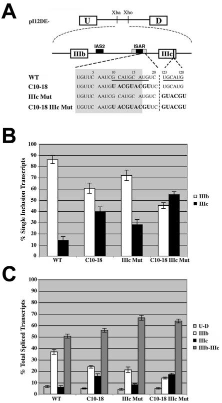
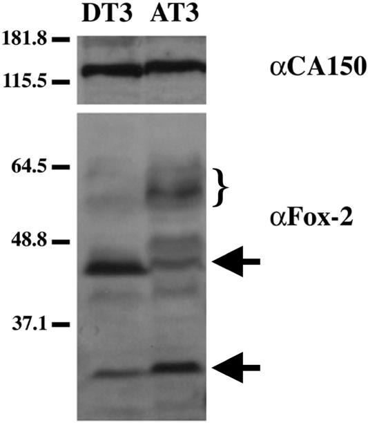
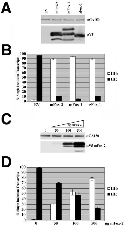
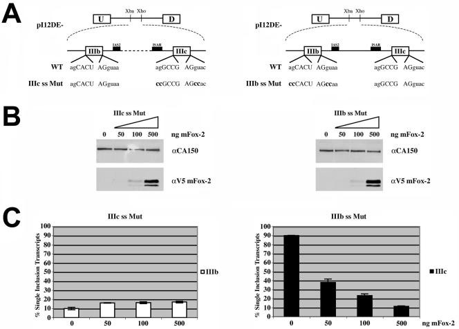
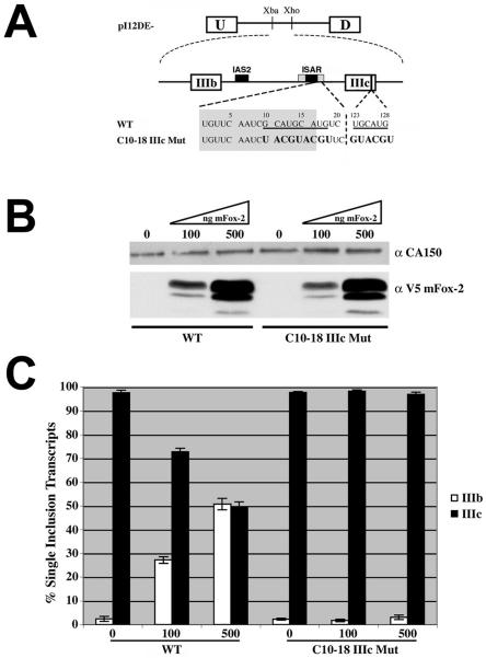
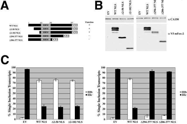
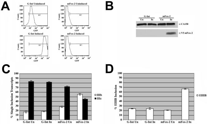
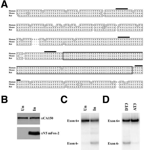
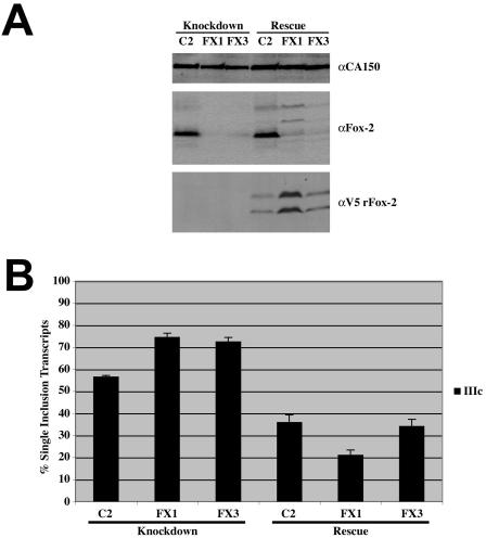
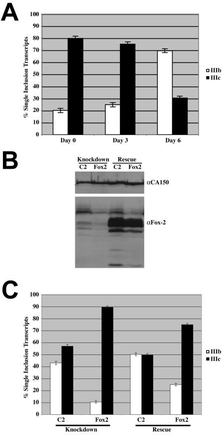
Alternative splicing of fibroblast growth factor receptor 2 (FGFR2) transcripts occurs in a cell-type-specific manner leading to the mutually exclusive use of exon IIIb in epithelia or exon IIIc in mesenchyme. Epithelial cell-specific exon choice is dependent on (U)GCAUG elements, which have been shown to bind Fox protein family members. In this paper we show that FGFR2 exon choice is regulated by (U)GCAUG elements and Fox protein family members. Fox-2 isoforms are differentially expressed in IIIb+ cells in comparison to IIIc+ cells, and expression of Fox-1 or Fox-2 in the latter led to a striking alteration in FGFR2 splice choice from IIIc to IIIb. This switch was absolutely dependent on the (U)GCAUG elements present in the FGFR2 pre-mRNA and required critical residues in the C-terminal region of Fox-2. Interestingly, Fox-2 expression led to skipping of exon 6 among endogenous Fox-2 transcripts and formation of an inactive Fox-2 isoform, which suggests that Fox-2 can regulate its own activity. Moreover, the repression of exon IIIc in IIIb+ cells was abrogated by interfering RNA-mediated knockdown of Fox-2. We also show that Fox-2 is critical for the FGFR2(IIIb)-to-FGFR2(IIIc) switch observed in T Rex-293 cells grown to overconfluency. Overconfluent T Rex-293 cells show molecular and morphological changes consistent with a mesenchymal-to-epithelial transition. If overconfluent cells are depleted of Fox-2, the switch from IIIc to IIIb is abrogated. The data in this paper place Fox-2 among critical regulators of gene expression during mesenchymal-epithelial transitions and demonstrate that this action of Fox-2 is mediated by mechanisms distinct from those described for other cases of Fox activity.


Figures

FIG. 1.
FIG. 1.
Intronic and exonic (U)GCAUG elements are critical for exon IIIb activation and exon IIIc repression in DT3 cells. (A) Schematic of minigenes used to test the importance of (U)GCAUG elements located downstream of ISAR core and in exon IIIc. Wild-type (U)GCAUG elements are underlined. Mutated nucleotides are indicated in boldface. Numbers above nucleotides represent base pair numbers downstream of ISAR core and within exon IIIc, respectively. IAS2 and ISAR core are represented as black boxes; ISAR core resides within the full ISAR element (represented as a gray box). Nucleotides in the gray box are located within ISAR. (B) The percent inclusion among single inclusion transcripts (U-IIIb-D and U-IIIc-D) produced by minigenes in panel A, which were stably integrated in DT3 cells, was determined by Invader RNA assay [e.g., % IIIb = 100 × number of U-IIIb-D transcripts/(number of U-IIIb-D transcripts + number of U-IIIc-D transcripts)]. (C) Quantification of all spliced products for transcripts from minigenes in panel A, which were stably integrated into DT3 cells, was determined by Invader RNA assay [e.g., % U-IIIb-D = 100 × number of U-IIIb-D transcripts/(number of U-D transcripts + number of U-IIIb-D transcripts + number of U-IIIc-D transcripts + number of U-IIIb-IIIc-D transcripts)].
FIG. 2.
FIG. 2.
Expression levels of Fox-2 isoforms differ between DT3 and AT3 cells. Western blot analysis was performed on total cellular protein isolated from DT3 and AT3 cells. The blot was probed for CA150 as a loading control and Fox-2 to detect the endogenous expression of Fox-2 proteins in AT3 and DT3 cells. The arrows indicate the presence of confirmed Fox-2 isoforms. The bracket indicates the presence of presumed Fox-2 isoforms. Numbers at left are molecular masses in kilodaltons.
FIG. 3.
FIG. 3.
zFox-1, mFox-1, and mFox-2 overexpression switches splicing pattern of FGFR2 minigene construct. (A) Western blot analysis was performed following transfection of 1 μg of V5-tagged mFox-2, mFox-1, or zFox-1 expression plasmids in HEK293T cells. The blot was probed for CA150 as a loading control and V5 to detect expression of mFox-2, mFox-1, and zFox-1. (B) Quantification of single inclusion transcripts by RT-PCR analysis of transiently transfected minigene with empty vector (EV), mFox-2, mFox-1, or zFox-1 expression plasmids. (C) Western blot analysis was performed following transfection of 0, 50, 100, and 500 ng of mFox-2 expression plasmid in HEK293T cells. The blot was probed for CA150 as a loading control and V5 to detect increasing expression of mFox-2. (D) Quantification of single inclusion transcripts by RT-PCR analysis of transiently transfected minigene with increasing amounts of mFox-2 expression plasmid.
FIG. 4.
FIG. 4.
mFox-2 overexpression activates exon IIIb and represses exon IIIc inclusion. (A) Schematic of minigenes used to test ability of mFox-2 expression to activate exon IIIb and repress exon IIIc independently of each other. (B) Western blot analysis was performed following transfection of increasing amounts of mFox-2 expression plasmid in HEK293T cells. The blot was probed for CA150 as a loading control and V5 to detect increasing amounts of mFox-2 expression. (C) Quantification of single inclusion transcripts by RT-PCR analysis of transiently transfected minigene reporters with increasing amounts of mFox-2 expression plasmid.
FIG. 5.
FIG. 5.
Intronic and exonic (U)GCAUG elements are critical for mFox-2-mediated FGFR2 exon IIIb activation and exon IIIc repression. (A) Schematic of minigenes used to test the role of (U)GCAUG elements in mFox-2-mediated exon IIIb activation and exon IIIc repression. Wild-type (U)GCAUG elements are underlined. Mutated nucleotides are indicated in boldface. Numbers above nucleotides represent base pair numbers downstream of ISAR core and within exon IIIc, respectively. IAS2 and ISAR core are represented as black boxes; ISAR core resides within the ISAR element (represented as a gray box). Nucleotides in the gray box reside within ISAR. (B) Western blot analysis was performed following transient transfection of increasing amounts of mFox-2 expression plasmid in HEK293T cells with WT and C10-18 IIIc Mut minigene reporters. The blot was probed for CA150 as a loading control and V5 to detect increasing expression of mFox-2. (C) Quantification of single inclusion transcripts by RT-PCR analysis of transiently transfected minigene reporters with increasing amounts of mFox-2 expression plasmid in HEK293T cells.
FIG. 6.
FIG. 6.
The last 82 amino acids of mFox-2 are required for switching FGFR2 exon choice. (A) Schematic of WT, amino-terminal deletion, and carboxy-terminal deletion proteins. The RRM is shown in gray. The NLS (N) and V5 epitope tags are located at the carboxy-terminal portion of the protein. Functionality of the protein was determined by the ability of the expressed protein to change the FGFR2 splicing pattern. (B) Western blot analysis was performed following transient transfection of WT NLS and mutant NLS expression plasmids in HEK293T cells. The blot was probed for CA150 as a loading control and V5 to detect expression of WT NLS and mutant NLS proteins. (C) Quantification of single inclusion transcripts by RT-PCR analysis of transiently transfected minigene with empty vector (EV), WT mFox-2 NLS, and each mutant construct.
FIG. 7.
FIG. 7.
mFox-2 induction switches endogenous FGFR2 splicing pattern and promotes exon EIIIB inclusion in endogenous fibronectin transcripts. Expression of GFP and mFox-2 in T Rex-293 cells was induced with 5 μg/ml of tetracycline. (A) FACS analysis was used to demonstrate the tetracycline-dependent induction of GFP in the T Rex-293 G-Int cell line. (B) Western blot analysis was used to demonstrate induction of V5-tagged mFox-2. The blot was probed for CA150 as a loading control and V5 to detect mFox-2. (C) Quantification of RT-PCR analysis for endogenous FGFR2 exon IIIb or exon IIIc inclusion of uninduced (Un) or induced (In) T Rex-293 G-Int and mFox-2 cell lines. (D) Quantification of RT-PCR analysis for endogenous fibronectin exon EIIIB inclusion of uninduced (Un) or induced (In) T Rex-293 G-Int and mFox-2 cell lines.
FIG. 8.
FIG. 8.
Induction of mFox-2 expression regulates alternative splicing of hFox-2 exon 6. Expression of mFox-2 in T Rex-293 cells was induced as described for Fig. 7. (A) ClustalW alignment of Fox-2 genomic sequence from human, mouse, and rat. TGCATG elements are indicated with black overlines, and exon 6 is enclosed with a box. (B) Western blot analysis was used to demonstrate induction of V5-tagged mFox-2. The blot was probed for CA150 as a loading control and V5 to detect mFox-2. (C) RT-PCR analysis of hFox-2 exon 6 inclusion. Induction of mFox-2 results in an increase in exon 6 transcripts. (D) RT-PCR analysis of rFox-2 exon 6 inclusion in DT3 and AT3 cells. DT3 cells contain more Fox-2 transcripts that are exon 6 compared to the AT3 cells.
FIG. 9.
FIG. 9.
RNAi-mediated Fox-2 knockdown allows for increased exon IIIc inclusion in DT3 cells. RNAi-resistant Fox-2 expression restores exon IIIc repression. (A) Western blot analysis was performed following transfection of siRNAs directed at no target (C2) or Fox-2 (FX1 and FX3). The blot was probed for CA150 as a loading control, Fox-2 to demonstrate knockdown and rescue of Fox-2 protein, and V5 to show expression of RNAi-resistant Fox-2 expression plasmid. (B) Quantification of single inclusion transcripts by RT-PCR analysis of transiently transfected pI12DE-IIIb ss Mut minigene reporter in cells treated with siRNAs and pTracer EF B (Knockdown) and cells treated with siRNAs and RNAi-resistant rFox-2 expression plasmids (Rescue).
FIG. 10.
FIG. 10.
Fox-2 is required for FGFR2 splicing switch during a mesenchymal-to-epithelial transition. (A) Quantification of RT-PCR analysis for endogenous FGFR2 exon IIIb or exon IIIc inclusion transcripts as T Rex-293 cells are grown to confluence. (B) Western blot analysis was performed following transfection of siRNAs directed at no target (C2) or Fox-2 (FX2). The blot was probed for CA150 as a loading control and Fox-2 to demonstrate knockdown of endogenous protein (Knockdown and Rescue) and expression of exogenous mFox-2 (Rescue). (C) Quantification of RT-PCR analysis for endogenous FGFR2 exon IIIb or exon IIIc inclusion transcripts in cells treated with siRNAs and pTracer EF B (Knockdown) and cells treated with siRNAs and the RNAi-resistant mFox-2 expression plasmid (Rescue).

References

    1. Baraniak, A. P., E. L. Lasda, E. J. Wagner, and M. A. Garcia-Blanco. 2003. A stem structure in fibroblast growth factor receptor 2 transcripts mediates cell-type-specific splicing by approximating intronic control elements. Mol. Cell. Biol. 23:9327-9337. - PMC - PubMed
    1. Brudno, M., M. S. Gelfand, S. Spengler, M. Zorn, I. Dubchak, and J. G. Conboy. 2001. Computational analysis of candidate intron regulatory elements for tissue-specific alternative pre-mRNA splicing. Nucleic Acids Res. 29:2338-2348. - PMC - PubMed
    1. Burns, R. C., T. J. Fairbanks, F. Sala, S. De Langhe, A. Mailleux, J. P. Thiery, C. Dickson, N. Itoh, D. Warburton, K. D. Anderson, and S. Bellusci. 2004. Requirement for fibroblast growth factor 10 or fibroblast growth factor receptor 2-IIIb signaling for cecal development in mouse. Dev. Biol. 265:61-74. - PubMed
    1. Carstens, R. P., J. V. Eaton, H. R. Krigman, P. J. Walther, and M. A. Garcia-Blanco. 1997. Alternative splicing of fibroblast growth factor receptor 2 (FGF-R2) in human prostate cancer. Oncogene 15:3059-3065. - PubMed
    1. Carstens, R. P., W. L. McKeehan, and M. A. Garcia-Blanco. 1998. An intronic sequence element mediates both activation and repression of rat fibroblast growth factor receptor 2 pre-mRNA splicing. Mol. Cell. Biol. 18:2205-2217. - PMC - PubMed

Publication types

MeSH terms

LinkOut - more resources