. 2006 Feb;26(4):1245-58.
doi: 10.1128/MCB.26.4.1245-1258.2006.

Developmental profile of H19 differentially methylated domain (DMD) deletion alleles reveals multiple roles of the DMD in regulating allelic expression and DNA methylation at the imprinted H19/Igf2 locus

Affiliations

Developmental profile of H19 differentially methylated domain (DMD) deletion alleles reveals multiple roles of the DMD in regulating allelic expression and DNA methylation at the imprinted H19/Igf2 locus

Joanne L Thorvaldsen et al. Mol Cell Biol. 2006 Feb.

Abstract

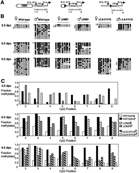
The differentially methylated domain (DMD) of the mouse H19 gene is a methylation-sensitive insulator that blocks access of the Igf2 gene to shared enhancers on the maternal allele and inactivates H19 expression on the methylated paternal allele. By analyzing H19 DMD deletion alleles H19DeltaDMD and H19Delta3.8kb-5'H19 in pre- and postimplantation embryos, we show that the DMD exhibits positive transcriptional activity and is required for H19 expression in blastocysts and full activation of H19 during subsequent development. We also show that the DMD is required to establish Igf2 imprinting by blocking access to shared enhancers when Igf2 monoallelic expression is initiated in postimplantation embryos and that the single remaining CTCF site of the H19DeltaDMD allele is unable to provide this function. Furthermore, our data demonstrate that sequence outside of the DMD can attract some paternal-allele-specific CpG methylation 5' of H19 in preimplantation embryos, although this methylation is not maintained during postimplantation in the absence of the DMD. Finally, we report a conditional allele floxing the 1.6-kb sequence deleted from the H19DeltaDMD allele and demonstrate that the DMD is required to maintain repression of the maternal Igf2 allele and the full activity of the paternal Igf2 allele in neonatal liver.


Figures

FIG. 1.
FIG. 1.
H19 expression from the mutant paternal alleles in pre- and postimplantation embryos. (A) Schematic of the H19ΔDMD and H19Δ3.8kb-5′H19 deletion alleles. The 2-kb DMD (unfilled box), the conserved 21-bp CTCF binding repeats (R1 to R4) (triangles), and H19 transcription unit (shaded box) are shown. Cross-hatched boxes represent sequence that was deleted from the alleles. The 1.6-kb KpnI-HindIII fragment, including repeats R2 to R4, was deleted from the H19ΔDMD allele, and the 3.8-kb SacI-XbaI fragment, including the entire DMD, was deleted from the H19Δ3.8kb-5′H19 allele. Arrows designate the genotyping primers G1 and G4 to G7. (B) H19 expression from the mutant paternal allele in the 3.5-dpc blastocysts, 6.5-dpc embryos, and 9.5-dpc embryos, yolk sacs, and placentas. Paternal-allele-specific expression (percent) was measured relative to expression from the wild-type maternal allele. Dark gray and light gray bars depict the results in the +/ΔDMD and +/Δ3.8kb-5′H19 samples, respectively. The average value for the samples assayed (N) is shown. As low levels of H19 expression were occasionally detected from the wild-type paternal allele (data not shown), only expression levels of >5% of that normally observed on the wild-type maternal allele were considered to be significant.
FIG. 2.
FIG. 2.
H19 expression from the mutant maternal alleles in pre- and postimplantation embryos. Results are shown for the 3.5-dpc blastocysts (A), 6.5-dpc embryos (B), and 9.5-dpc embryos, yolk sacs, and placentas (C). In each panel, H19 RNA relative to GAPDH (Gapd) RNA for wild-type and mutant littermates is presented graphically. Error bars reflect results from three experiments. A phosphorimage of the H19 and GAPDH RT-PCR products from one representative experiment is presented below each graph. Black, dark gray, and light gray bars depict the results in the +/+, ΔDMD/+, and Δ3.8kb-5′H19/+ samples, respectively.
FIG. 3.
FIG. 3.
Igf2 expression from the mutant maternal alleles in postimplantation embryos. (A) Igf2 exon 6 Tsp509I polymorphism. Schematic of the Tsp509I-digested Igf2 RT-PCR products derived from the wild-type B6 (+), H19ΔDMD and H19Δ3.8kb-5′H19 (Δ), and Cast (C) alleles. (B) Allelic analysis of all Igf2 transcripts. The genotype of the sample assayed is indicated above each lane (maternal/paternal). To the right of each panel, the undigested (U) and digested (+, Δ, and C) Igf2 RT-PCR products are noted. The presence (+) and absence (−) of Tsp509I and the tissue assayed are presented below each lane of the panels. In 6.5-dpc embryos (EM, top panel), Igf2 expression was measured in embryos inheriting the wild-type (+) or deletion allele from their mother or father. The marker lane (M) contains MspI-digested pBR322 DNA. The bottom panel shows Igf2 expression in 9.5-dpc placenta, embryo, and yolk sac (PL, EM, and YS) and in Cast (C) and B6 (+) neonatal liver. In ΔDMD/C and Δ3.8kb-5′H19/C 9.5-dpc placenta, embryo, and yolk sac, the ratio of maternally to paternally derived Igf2 ranged from 1.13 to 1.41 (PL), 0.96 to 1.15 (EM), and 1.06 to 1.18 (YS), respectively. There was no significant difference in Igf2 expression between ΔDMD/C and Δ3.8kb-5′H19/C samples. For all 6.5- and 9.5-dpc samples assayed, no PCR product was detected in the absence of the RT enzyme (data not shown). (C) Igf2 exon μ1 BanI polymorphism. The schematic shows the BanI-digested Igf2 RT-PCR products expressed from the wild-type B6 (+), H19ΔDMD and H19Δ3.8kb-5′H19 (Δ), and Cast (C) alleles. (D) Allelic Igf2 expression initiated from the Igf2 placenta-specific promoter P0. See the legend for panel B for panel description.
FIG. 4.
FIG. 4.
Methylation pattern of the 5′ DMD sequence on the ΔDMD alleles in pre- and postimplantation embryos. (A) Schematic of the wild-type and H19ΔDMD regions analyzed by the bisulfite mutagenesis and sequencing assay. Sixteen and seven CpG dinucleotides were assayed on the wild-type and H19ΔDMD alleles, respectively. The seven CpG dinucleotides at the 5′ DMD region (gray bar over open circles) are common to both the wild-type and deletion alleles. The H19ΔDMD CpG dinucleotides within the 5′ DMD sequence (1 to 7) are depicted. Above each allele are the primers (B1 to B4, B10, and B11) used to amplify the assayed regions. See Materials and Methods for location of primers relative to the H19 transcription start site. (B) Methylation status of the individual maternal and paternal DNA strands of the wild-type and H19ΔDMD alleles in 3.5-dpc, 6.5-dpc, and 9.5-dpc embryos. Methylated and unmethylated cytosines are represented as filled and open circles, respectively. An absent circle indicates the residue was not assayed. Profiles observed more than once are indicated to the left of each strand. On the H19ΔDMD alleles, the CpG dinucleotides within the loxP/vector sequence and sequence downstream of the deleted DMD sequence were also methylated exclusively on the paternal allele in the blastocysts and hypermethylated on both parental alleles in postimplantation embryos (data not shown). (C) Summary of the methylation profiles of the seven 5′ DMD CpG dinucleotides common to the wild-type and H19ΔDMD alleles in 3.5-dpc, 6.5-dpc, and 9.5-dpc embryos. White and black bars represent the fractions of methylated cytosines on the maternal and paternal wild-type alleles, respectively. Gray cross-hatched and gray solid bars represent the fractions of methylated cytosines on the maternal and paternal H19ΔDMD alleles, respectively.
FIG. 5.
FIG. 5.
Methylation pattern of the promoter proximal (PP) region on the wild-type and H19ΔDMD and H19Δ3.8kb-5′H19 alleles in pre- and postimplantation embryos. (A) Schematic of the wild-type and H19ΔDMD and H19Δ3.8-5′H19 regions analyzed by the bisulfite mutagenesis and sequencing assay. The nine CpG dinucleotides spanning the PP region were assayed on the wild-type and H19ΔDMD alleles. On the H19Δ3.8-5′H19 allele, as specified beneath the schematic (gray bars), the eight remaining PP CpGs (2-9) were assayed. The primers (B12-B16) used to amplify the mutagenized DNA are shown above each allele. See Materials and Methods for location of the primers relative to the H19 transcription start site. (B) The methylation status of the individual maternal and paternal DNA strands of the wild-type, H19ΔDMD and H19Δ3.8-5′H19 alleles in 3.5-dpc, 6.5-dpc and 9.5-dpc embryos. See Fig. 4 legend for details. On the H19Δ3.8-5′H19 alleles, the CpG dinucleotides within the loxP/vector sequence and sequence upstream of the deleted DMD sequence were also methylated exclusively on the paternal allele in sperm and blastocysts and hypermethylated on both parental alleles in postimplantation embryos (data not shown). (C) Summary of the methylation profiles of the eight PP CpG dinucleotides common to the wild-type, H19ΔDMD and H19Δ3.8-5′H19 alleles in 3.5-dpc, 6.5-dpc and 9.5-dpc embryos. White and black bars represent the fraction of methylated cytosines on the maternal and paternal wild-type alleles, respectively. Gray crosshatched and gray bars represent the fraction of methylated cytosines on the maternal and paternal H19ΔDMD alleles, respectively. Black crosshatched and light gray bars represent the fraction of methylated cytosines on the maternal and paternal H19Δ3.8-5′H19 alleles, respectively.
FIG. 6.
FIG. 6.
Conditional deletion of the 1.6-kb DMD sequence in neonatal liver. (A) Generation of the conditional H19lxDMDallele. Depicted from top to bottom are the wild-type H19 locus, targeting vector lxDMD, H19lxDMDneo targeted allele, H19lxDMD conditional allele after removal of the PGK-neo cassette, and the H19lxΔDMD deletion allele after AlbCre-mediated excision of the DMD in liver. Restriction site locations (in kb) are relative to the start of H19 transcription. Probes A (1.7 kb EcoRV-EcoRI fragment), B (0.75 kb BamHI-StuI fragment), and C (0.9 kb SacI-KpnI fragment) were used to identify clones with the correctly targeted and excised alleles. The DMD and the H19 exons are boxed. Vector lxDMD includes the DMD and a PGK-neo cassette (neo) (boxed), which are both flanked by loxP sites (wide arrowheads), flanking H19 DNA (thin line), and pBKSII (thick line). Primers (G1, G2, G3, G5, and G7) used to identify targeted and excised alleles are below lxDMD. (B) Southern analysis of the H19lxDMDneo, H19lxDMD, and H19lxΔDMD alleles. Probes A and B were hybridized to EcoRV- and StuI-digested DNA, respectively, to detect the parental E14 (lane 1) and targeted H19lxDMDneo ES cell clones (lane 2). Probe C was hybridized to SacI-digested liver DNA (with or without HhaI for methylation analysis) from 5-day and 10-day progeny of reciprocal crosses between H19lxDMD/+ and C7 AlbCre/+ mice (in lanes 1 to 5 H19lxDMD/+ was the mother; in lanes 6 to 10, H19lxDMD/+ was the father). The Cast (C, 1.6 kb), B6 (B, 3.8kb), H19lxDMD (lx, 3.9 kb) and H19lxΔDMD (lxΔ, 2.3 kb) SacI fragments are indicated. In the presence of AlbCreTg (lanes 3, 4, and 7 to 11), the excision allele H19lxΔDMD was detected in 40% and 60 to 70% of liver DNA from 5-day and 10-day progeny, respectively. The 5′ DMD remained hypomethylated on the maternal DMD deletion allele (lanes 8 and 9) and hypermethlyated on the paternal DMD deletion allele (lanes 10 and 11). (C) Igf2 and H19 expression in neonatal livers from mice inheriting the maternal H19lxΔDMD allele. Allele-specific Igf2 expression (top panel) and H19 expression relative to rpL32 (bottom panel) was measured in liver RNA from 5-day-old progeny of a mating between H19lxDMD/+ and AlbCreTgCAST7 mice. The presence AlbCreTg (Alb-Cre) is noted below each panel in sections C and D. The ratio of maternally to paternally derived Igf2 RNA is 0.01, 0.01, 0.02, 0.02, 0.02, 0.01, 0.24, 0.28 and 0.23 in lanes 1 to 9, respectively. The ratio of H19 to rpL32 RNA is 3.18, 3.96, 3.86 and 2.19 in lanes 1 to 4, respectively. (D) H19 and Igf2 expression in mice with the paternal H19lxΔDMD allele in neonatal liver. Allele-specific H19 expression (top panel) and Igf2 expression relative to rpL32 (bottom panel) was measured in Li RNA from 5-day-old progeny of a mating between AlbCreTgCAST7 and H19lxDMD/+ mice. The ratio of paternally to maternally derived H19 RNA is 0.01, 0.02, 0.06, 0.07, 0.12, and 0.13 in lanes 1 to 6, respectively. The ratio of Igf2 to rpL32 RNA is 3.97, 5.17, 2.26, and 1.94 in lanes 1 to 4, respectively. The average Igf2/rpl32 RNA levels of four Cast/B6 and four Cast/lxΔDMD neonatal livers were 3.93 and 1.69, respectively. The difference was statistically significant (P = 0.04).

Similar articles

Cited by

References

    1. Auffray, C., and F. Rougeon. 1980. Purification of mouse immunoglobulin heavy-chain messenger RNAs from total myeloma tumor RNA. Eur. J. Biochem. 107:303-314. - PubMed
    1. Bartolomei, M. S., A. L. Webber, M. E. Brunkow, and S. M. Tilghman. 1993. Epigenetic mechanisms underlying the imprinting of the mouse H19 gene. Genes Dev. 7:1663-1673. - PubMed
    1. Bell, A. C., and G. Felsenfeld. 2000. Methylation of a CTCF-dependent boundary controls imprinted expression of the Igf2 gene. Nature 405:482-485. - PubMed
    1. Cho, H., J. Mu, J. K. Kim, J. L. Thorvaldsen, Q. Chu, E. B. Crenshaw III, K. H. Kaestner, M. S. Bartolomei, G. I. Shulman, and M. J. Birnbaum. 2001. Insulin resistance and a diabetes mellitus-like syndrome in mice lacking the protein kinase Akt2 (PKB beta). Science 292:1728-1731. - PubMed
    1. Cranston, M. J., T. L. Spinka, D. A. Elson, and M. S. Bartolomei. 2001. Elucidation of the minimal sequence required to imprint H19 transgenes. Genomics 73:98-107. - PubMed

Publication types

LinkOut - more resources