. 2006 Feb;26(4):1333-46.
doi: 10.1128/MCB.26.4.1333-1346.2006.

Splicing of a critical exon of human Survival Motor Neuron is regulated by a unique silencer element located in the last intron

Affiliations

Splicing of a critical exon of human Survival Motor Neuron is regulated by a unique silencer element located in the last intron

Nirmal K Singh et al. Mol Cell Biol. 2006 Feb.

Abstract

Humans have two nearly identical copies of the Survival Motor Neuron (SMN) gene, SMN1 and SMN2. In spinal muscular atrophy (SMA), SMN2 is not able to compensate for the loss of SMN1 due to exclusion of exon 7. Here we describe a novel inhibitory element located immediately downstream of the 5' splice site in intron 7. We call this element intronic splicing silencer N1 (ISS-N1). Deletion of ISS-N1 promoted exon 7 inclusion in mRNAs derived from the SMN2 minigene. Underlining the dominant role of ISS-N1 in exon 7 skipping, abrogation of a number of positive cis elements was tolerated when ISS-N1 was deleted. Confirming the silencer function of ISS-N1, an antisense oligonucleotide against ISS-N1 restored exon 7 inclusion in mRNAs derived from the SMN2 minigene or from endogenous SMN2. Consistently, this oligonucleotide increased the levels of SMN protein in SMA patient-derived cells that carry only the SMN2 gene. Our findings underscore for the first time the profound impact of an evolutionarily nonconserved intronic element on SMN2 exon 7 splicing. Considering that oligonucleotides annealing to intronic sequences do not interfere with exon-junction complex formation or mRNA transport and translation, ISS-N1 provides a very specific and efficient therapeutic target for antisense oligonucleotide-mediated correction of SMN2 splicing in SMA.


Figures

FIG. 1.
FIG. 1.
Model of cis elements that regulate splicing of exon 7 of human SMN. Uppercase letters represent exon 7 sequences. Lowercase letters represent intronic sequences. The star represents position 6, where a C is replaced by a U (C6U) in SMN2 exon 7. The U1 snRNA binding site that spans the first six nucleotides of intron 7 is shaded. Based on analysis of the entire exon 7 (51), three major cis elements (Exinct, Conserved tract, and 3′-Cluster) are shown. The putative binding sites of SF2/ASF (5) and hnRNP-A1 (24) fall within the inhibitory cis element Exinct. The binding site of Tra2-β1 (18) falls within the stimulatory cis element Conserved tract.
FIG. 2.
FIG. 2.
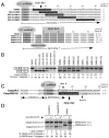
Effects of intronic deletions downstream of the 5′ ss of exon 7 of SMN2. (A) Intronic sequences of SMN2 deletion mutants. Nucleotide numbering starts from the beginning of intron 7. Deletions are shown as dashed lines. The ISS-N1 site is shaded. Nucleotides involved in base pairing with U1 snRNA are in bold and shaded. Numbers in mutants' names represent the positions at which deletions were made. (B) In vivo splicing pattern of SMN2 deletion mutants shown in panel A. The upper band corresponds to a fully spliced product that includes exon 7; the lower band corresponds to an exon 7-skipped product. The percentage of exon 7 skipping was calculated from the total value of exon 7-included and exon 7-skipped products. Abbreviations E6, E7, and E8 stand for exon 6, exon 7, and exon 8, respectively. (C) In vivo splicing pattern of SMN2ΔISS-N1 (ISS-N1-deleted mutant) in different cell lines. Of note, SMN2ΔISS-N1 is the same construct as the N1Δ10-24 mutant in panel A. Cell lines used were Neuro-2a (mouse brain neuroblastoma, lanes 1 to 3), NSC-34 (mouse motor neuron like, lanes 4 to 6), SK-N-SH (human neuroblastoma, lanes 7 to 9), P-19 (mouse embryonic teratocarcinoma, lanes 10 to 12), and HEK-293 (lanes 13 to 15). Spliced products are the same as those indicated in panel B. NA, not applicable.
FIG. 3.
FIG. 3.
Evolutionary significance of ISS-N1. (A) Alignment of the first 42 nucleotides of human and mouse introns 7, including the ISS-N1 region. Numbering starts from the beginning of intron 7. ISS-N1 is shaded. Nucleotides involved in base pairing with U1 snRNA are in bold and shaded. In the top two lines, positions homologous between the human and mouse sequences are shaded in black. In the bottom 10 lines, intronic sequences of SMN2 mutants (SMN2/MS1 through SMN2/MS10) are shown with mouse nucleotides written in lowercase letters and shaded in black. (B) In vivo splicing pattern of SMN2 mutants shown in panel A. Spliced products are the same as those indicated in Fig. 2B. NA, not applicable.
FIG. 4.
FIG. 4.
Effects of antisense oligonucleotides on splicing of different minigenes. (A) Diagrammatic representation of intron 7 regions targeted by antisense oligonucleotides Anti-N1, Anti-N1+10, Anti-N1+20, and Anti-N1+30. Numbering starts from the beginning of intron 7. The ISS-N1 region is shaded. Of the four oligonucleotides shown, only Anti-N1 fully sequestered ISS-N1. (B) In vivo splicing pattern of the SMN2 minigene in the presence of antisense oligonucleotides. Spliced products are the same as those indicated in Fig. 2B. (C) In vivo splicing pattern of different minigenes in the presence of Anti-N1. In every lane, the upper band represents the exon-included and the lower band represents the exon-excluded products. In CFTR, apoA-II, Fas, and Casp3, the major bands represent the exon-included products. In Tau and Fas (U-20C), the major bands represent the exon-excluded products. For detection of spliced products in lanes 1 to 6, 9, and 10, cells were harvested 40 h after transfection (for other lanes, see Materials and Methods). The values on the left are sizes in base pairs. wt, wild type; NA, not applicable.
FIG. 5.
FIG. 5.
Effect of base pairing between Anti-N1 and ISS-N1 on the efficiency of SMN2 exon 7 inclusion. (A) Intron 7 sequences of SMN2 and mutants with substitutions in the ISS-N1 region. Numbering starts from the beginning of intron 7. The ISS-N1 region is shaded in gray. Nucleotides involved in base pairing with U1 snRNA are in bold and shaded. Note that intronic mutations (shaded in black) abrogate base pairing between Anti-N1 and ISS-N1. (B) Sequences of Anti-N1 and Anti-I7-25 oligonucleotides. Differences are shaded in black. Note that Anti-I7-25 restores base pairing with the ISS-N1 region in mutant SMN2/I7-25. (C) In vivo splicing pattern of mutants shown in panel A. Plasmid DNA (0.1 μg) was transfected alone or cotransfected with 50 nM Anti-N1 or Anti-I7-25 oligonucleotide. Spliced products are the same as those indicated in Fig. 2B.
FIG. 6.
FIG. 6.
Relative significance of exon 7 cis elements vis-à-vis ISS-N1. (A) Diagrammatic representation of several cis elements involved in regulation of exon 7 splicing (not to scale). 1G, Tra2-β1 (Tra2-ESE), CT (Conserved tract), and element 2 represent positive elements (+). ISS-N1 is a negative element (−). (B) In vivo splicing pattern of SMN1 mutants in which deletion of ISS-N1 (ΔISS-N1) was combined with abrogation of a given positive cis element. Spliced products are the same as those indicated in Fig. 2B. Abr-E2 represents abrogation of element 2 by a triple substitution G69C/U70A/U71A in intron 7 (40), Abr-Tra2 represents abrogation of Tra2-ESE by a 25U26U mutation in exon 7 (18), 1U mutation represents abrogation of a cis element at the first position (51), and Abr-CT represents abrogation of Conserved tract by a 36U37U mutation in exon 7 (51).
FIG. 7.
FIG. 7.
Portability of ISS-N1. (A, top) Location of ISS-N1 within SMN2 intron 7 with respect to the 5′ ss. ISS-N1 was inserted at different locations within intron 7 of SMN2ΔISS-N1. (A, bottom) Intronic sequences of SMN2 mutants with five-nucleotide-long insertions immediately upstream of ISS-N1. Nucleotide positions and types of insertions are indicated. Numbering starts from the beginning of intron 7. ISS-N1 is shaded. Nucleotides involved in base pairing with U1 snRNA are in bold and shaded. (B) In vivo splicing pattern of mutants shown in panel A. Spliced products are the same as those indicated in Fig. 2B. (C) Insertion of ISS-N1 in a heterologous context. For insertion of ISS-N1, an AvrII restriction site was first inserted downstream of exon 6 of the Casp3 minigene. (D) In vivo splicing pattern of mutants shown in panel C. The splicing pattern was determined in the absence and presence of an antisense oligonucleotide (Anti-ISS-N1/15) that fully sequesters ISS-N1. In the absence of Anti-ISS-N1/15, the Casp3ISS-N1 mutant increases exclusion of Casp3 exon 6 (compare lane 3 with lane 4). NA, not applicable.
FIG. 8.
FIG. 8.
Effect of Anti-N1 on the splicing of endogenous genes. (A) Splicing of endogenous SMN2 after SMA fibroblasts (GM03813) were transfected with 5 nM oligonucleotides. Total RNA was collected 25 h after transfection. (B) Specificity of the effect of Aniti-N1 on the splicing pattern of other exons in SMA fibroblasts (GM03813). The sizes of the expected spliced products are indicated on the left. The same RNA as that used in panel A (lanes 2 and 3) was used for analysis. The 662-bp bands in lanes 1 and 2 represent the SMN exon 3-included product, and the 387-bp bands in lanes 3 and 4 represent the SMN exon 5-included product. The 440-bp bands in lanes 5 and 6 represent transcripts that exclude exon 2b but include exon 3 of Survivin (34). The 830-bp bands in lanes 7 and 8 represent transcripts that include exons 29 and 30 of NF1 (48). The 686-bp bands in lanes 9 and 10 represent transcripts that produce a Tra2-β1 spliced variant of Tra2 (8). The 474-bp bands in lanes 11 and 12 represent the Caspase 3 exon 6-included product (21). The 300-bp bands in lanes 13 and 14 represent a Bcl-xL spliced variant of Bcl-x (37). (C) Effect of Aniti-N1 on the level of SMN protein. Western blotting was performed to detect SMN in SMA fibroblasts (GM03813) transfected with 5 nM (lane 1) and 15 nM (lane 3) Anti-N1. GM03813 cells transfected with control oligonucleotide Scramble20 (lanes 2 and 4) and mock-transfected GM03813 cells (lane 5) or AG06814 cells (normal fibroblasts) (lane 6) were used as controls. For detection of SMN, cells were harvested 72 h after transfection. We used α-tubulin as a loading control. The values on the left are sizes in base pairs.

References

    1. Bentley, D. L. 2005. Rules of engagement: co-transcriptional recruitment of pre-mRNA processing factors. Curr. Opin. Cell Biol. 17:251-256. - PubMed
    1. Black, D. L. 2003. Mechanisms of alternative pre-messenger RNA splicing. Annu. Rev. Biochem. 72:291-336. - PubMed
    1. Buratti, E., and F. E. Baralle. 2004. Influence of RNA structure on the pre-mRNA splicing process. Mol. Cell. Biol. 24:10505-10514. - PMC - PubMed
    1. Buratti, E., and F. E. Baralle. 2005. Another step forward for SELEXive splicing. Trends Mol. Med. 11:5-9. - PubMed
    1. Cartegni, L., and A. R. Krainer. 2002. Disruption of an SF2/ASF-dependent exonic splicing enhancer in SMN2 causes spinal muscular atrophy in the absence of SMN1. Nat. Genet. 30:377-384. - PubMed

Publication types

MeSH terms

LinkOut - more resources