. 2006 Feb;26(4):1434-44.
doi: 10.1128/MCB.26.4.1434-1444.2006.

Efficient and specific targeting of Polycomb group proteins requires cooperative interaction between Grainyhead and Pleiohomeotic

Affiliations

Efficient and specific targeting of Polycomb group proteins requires cooperative interaction between Grainyhead and Pleiohomeotic

András Blastyák et al. Mol Cell Biol. 2006 Feb.

Abstract

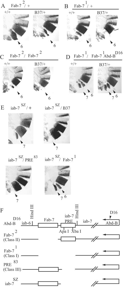
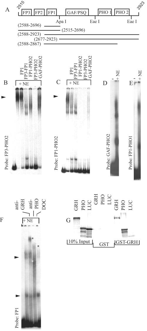
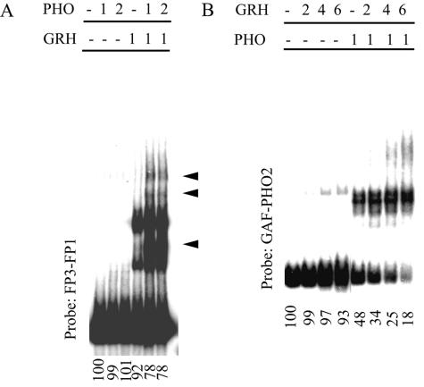
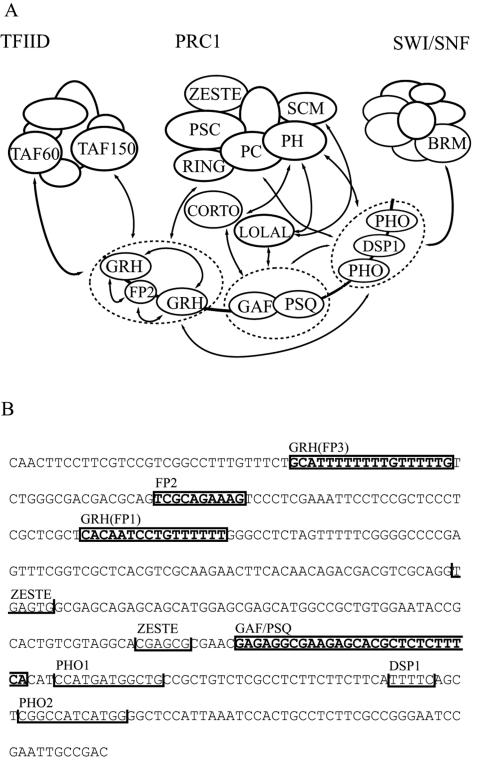
Specific targeting of the protein complexes formed by the Polycomb group of proteins is critically required to maintain the inactive state of a group of developmentally regulated genes. Although the role of DNA binding proteins in this process has been well established, it is still not understood how these proteins target the Polycomb complexes specifically to their response elements. Here we show that the grainyhead gene, which encodes a DNA binding protein, interacts with one such Polycomb response element of the bithorax complex. Grainyhead binds to this element in vitro. Moreover, grainyhead interacts genetically with pleiohomeotic in a transgene-based, pairing-dependent silencing assay. Grainyhead also interacts with Pleiohomeotic in vitro, which facilitates the binding of both proteins to their respective target DNAs. Such interactions between two DNA binding proteins could provide the basis for the cooperative assembly of a nucleoprotein complex formed in vitro. Based on these results and the available data, we propose that the role of DNA binding proteins in Polycomb group-dependent silencing could be described by a model very similar to that of an enhanceosome, wherein the unique arrangement of protein-protein interaction modules exposed by the cooperatively interacting DNA binding proteins provides targeting specificity.


Figures

FIG. 1.
FIG. 1.
grainyhead interacts with the iab-7 PRE. (A to D) Abdominal cuticle preparations of adult male flies carrying internal BX-C deletions in the wild-type (left side of each panel) or grhB37 mutant (right side of each panel) background. The BX-C deletions are Fab-72/+ (A), Fab-71/+ (B), Fab-72/Fab-72 (C), and Fab-71/Fab-71 Abd-BD16 (D). The numbered arrows show the relevant abdominal segments. A7 normally does not have a visible tergite, and the size reduction of the A6 tergite indicates a partial transformation toward an A7 identity of this segment. Increasing reduction of the tergite size indicates increasingly stronger transformation. (E) grainyhead interaction with the iab-7SZ deletion can be also detected even though the boundary function remains unaffected by this deletion. Note that the phenotype of grhB37/iab-7SZ transheterozygous flies is very similar to that of class III/iab-7SZ flies, while it is markedly different from that of the Fab-71/iab-7SZ combination. (F) Schematic representation of BX-C mutants. For exact molecular coordinates, see reference . The Fab-7 boundary and the iab-7 PRE are represented by boxes.
FIG. 2.
FIG. 2.
grainyhead interacts genetically with Sex combs extra (Sce). The first, second, and third legs of wild-type (A to C) and grhB37/+ Sce1/+ (D to F) male flies are shown. Arrows point to sex combs, whose presence on the second and third legs indicate partial transformation of the respective thoracic segments (T2 and T3) into a T1 identity, which is a characteristic phenotype of certain Pc-G alleles and allele combinations (8). The average number of legs having at least one sex comb tooth is 2.2 in Sce1 heterozygous male flies (n = 177) and 5.4 in grhB37/+ Sce1/+ transheterozygous males (n = 220). We have never observed ectopic sex combs on grh heterozygous flies.
FIG. 3.
FIG. 3.
DNase I footprinting demonstrates GRH binding to the iab-7 PRE. The probe fragment used here was amplified by the P1 and P5 primers (see Materials and Methods) and labeled at the proximal end (A and B) or at the distal end (C). Bacterially expressed GRH was used as the protein source for the experiment shown in panel A, and nuclear extract (NE) was used for the experiments shown in panels B and C, where the resulting footprints (FP) are numbered. In panel B, instead of clear protection of the sequence, protein binding to FP1 is indicated only by the increased DNase I sensitivity around the FP1 site. Note that the marked changes in the DNase I digestion pattern between FP1-FP2 and FP2-FP3, indicated by brackets on both strands, are highly compatible with the short-range looping model described in the text. FP2 corresponds to the sequence TCGCAGAAAG. (D) Anti-GRH antibody recognizes both complexes formed on a single FP1 site in a gel shift assay. GRH-specific shifts are marked by arrowheads throughout. Supershifts are indicated by asterisks. (E) In a gel shift experiment, bacterially expressed GRH binds to a wild-type (w.t.) FP3 oligonucleotide (CTGCATTTTTTTTGTTTTTGTCT) but not to the mutated (mut.) sequence (CTGCATTTTTTTTCACTTTGTCT[the mutation is underlined]). Gel shifts demonstrate cooperative binding of GRH. (F) GRH binding to the FP3 motif in a gel shift assay can be deduced from the competition of the marked shifts by nonoverlapping GRH binding site FP1 (the applied competitor fragments are referred to by their proximal and distal binding sites and are indicated above the lanes). (G) The upper GRH-specific shift detected with the FP3-FP2 probe is essentially absent from the FP3 probe, which indicates context-dependent, cooperative interaction between FP3 and FP2. (H) Another level of cooperativity is suggested by the experiment with the FP3-FP1 probe, since the formation of the single complex on this probe cannot be interpreted as the sum of the pattern of binding to its subfragments (compare with panels D, F, and G). Moreover, subfragments of the probe could not compete as efficiently for binding as the large fragment itself. (I) Schematic representation of the subfragments used as probes and as competitors in the gel shift experiments. Binding sites are represented by boxes. Molecular coordinates are shown in parentheses according to EMBL X78983.
FIG. 4.
FIG. 4.
Transgenic constructs carrying iab-7 PRE fragments interact genetically with grh. (A) A typical homozygous type I transformant line that responds to grhB37. Note that in a wild-type background the reporter gene is completely repressed. Such strong silencing was observed in 7 out of the 22 established PS lines. (B) In the case of PS lines carrying a type II construct, the reporter gene is never completely silenced. (C) Type III transformant lines show no signs of pairing sensitivity. (D) In this type I transformant line, neither grhB37 nor pho1 caused a dramatic change in the eye color of the homozygous transgene. However, in the double-mutant background significant derepression of the reporter gene is evident, indicating synergism between grh and pho. (E) Schematic representation of the transgenic constructs. The boxes represent binding sites for DNA binding proteins. Molecular coordinates are given according to EMBL X78983. The number of PS versus homozygous (hom.) viable lines obtained for each construct is indicated on the right.
FIG. 5.
FIG. 5.
A highly cooperative nucleoprotein complex is formed on the iab-7 PRE in vitro. (A) Schematic representation of probes and subfragments. Binding sites for DNA binding proteins are represented by boxes. Molecular coordinates are given in parentheses according to EMBL X78983. These subfragments are referred to by their proximal and distal binding sites throughout. (B) When the FP3-PHO2 template is used in gel shift experiments, almost all of the probe is bound and the resulting complex is highly resistant to competition either by the single binding site FP1 or even by the GAF/PSQ-PHO2 subfragment, indicating cooperative assembly of a protein complex. (C) Signs of cooperativity are still evident, although to a lesser extent, when the FP1-PHO2 template is used. Further truncation of this probe to remove the FP1 or the PHO2 site results in loss of cooperative binding (panels D and E, respectively). (F) The protein complex formed on the FP1 GRH site is supershifted by both anti-GRH and anti PHO, indicating that GRH and PHO are members of the same protein complex. Supershifts are indicated by asterisks. The effect of deoxycholate (DOC) is also shown; this detergent eliminated the upper, but not the lower, shift at a concentration of 0.1%, indicating that the upper shift is the result of a detergent-sensitive protein-protein interaction. (G) In a GST pulldown experiment, PHO interacts with GRH. GRH homodimerization (4, 59) was used as a positive and luciferase (LUC) as a negative control for binding. PHO binds to the GST-GRH matrix but not to GST alone.
FIG. 6.
FIG. 6.
Mutual enhancement of DNA binding between PHO and GRH. (A) A constant amount of GRH is titrated in the presence of increasing amounts of PHO. The probe fragments used here cover the FP3 and FP1 sites but lack the PHO sites (see also Fig. 4E). GRH-PHO complexes are marked by arrowheads. (B) Binding of a constant amount of PHO in the presence of increasing amounts of GRH to a probe which contains only the PHO sites without the FP3 and FP1 sites (see Fig. 4E). A discrete PHO-GRH complex was not detected. Nevertheless, cooperativity can be deduced from the decreasing amount of free DNA, which faithfully reflects the original equilibrium situation even if the specific complexes dissociate during electrophoresis. The amount of free DNA quantified by a PhosphorImager is shown below each lane as a percentage of the input DNA. The amounts of GRH and PHO are marked as relative units above the lanes.
FIG. 7.
FIG. 7.
(A) Model of PRC1 targeting to iab-7 PRE. For simplicity, similar DNA binding proteins are circled and treated as single targeting domains. Cooperative interactions between DNA binding proteins and subunits of PRC1 are indicated by arrows. These interactions are described in the text and in references , , , , and . Based on the available data, the interaction of GAF with PRC1 subunits requires the adaptor proteins LOLA-LIKE and CORTO (38, 48). The possible roles of other proteins, such as RYBP and CtBP, should also be considered (3, 21, 54). The putative GAF-PHO interaction would require a nucleosomal context (35). TFIID can interact with GRH (11), whereas SWI/SNF can interact with PHO (40). Therefore, it would be conceivable that these two complexes can also be recruited to the iab-7 PRE. According to our model, however, stable recruitment of TFIID and SWI/SNF is unlikely to occur because their binding is not supported by other interacting modules in the context of the iab-7 PRE. (B) Locations of binding sites for regulatory proteins within the largest iab-7 PRE fragment used in this study are shown. Closed boxes indicate footprinted sequences. The boundaries of the FP3, FP2, and FP1 footprints are derived from experiments with nuclear extract, and the FP1 site was also verified by using purified protein. Protection of the GAF/PSQ motif was detected with nuclear extract (data not shown; 39). Consensus sites for the PHO, ZESTE, and DSP1 proteins are indicated by open boxes (14, 15, 39).

Similar articles

Cited by

References

    1. Americo, J., M. Whiteley, J. L. Brown, M. Fujioka, J. B. Jaynes, and J. A. Kassis. 2002. A complex array of DNA-binding proteins required for pairing-sensitive silencing by a polycomb group response element from the Drosophila engrailed gene. Genetics 160:1561-1571. - PMC - PubMed
    1. Armstrong, J. A., O. Papoulas, G. Daubresse, A. S. Sperling, J. T. Lis, M. P. Scott, and J. W. Tamkun. 2002. The Drosophila BRM complex facilitates global transcription by RNA polymerase II. EMBO J. 21:5245-5254. - PMC - PubMed
    1. Atchison, L., A. Ghias, F. Wilkinson, N. Bonini, and M. L. Atchison. 2003. Transcription factor YY1 functions as a PcG protein in vivo. EMBO J. 22:1347-1358. - PMC - PubMed
    1. Attardi, L. D., and R. Tjian. 1993. Drosophila tissue-specific transcription factor NTF-1 contains a novel isoleucine-rich activation motif. Genes Dev. 7:1341-1353. - PubMed
    1. Bray, S. J., B. Burke, N. H. Brown, and J. Hirsh. 1989. Embryonic expression pattern of a family of Drosophila proteins that interact with a central nervous system regulatory element. Genes Dev. 3:1130-1145. - PubMed

Publication types

MeSH terms

LinkOut - more resources