. 2006 Feb;26(4):1480-95.
doi: 10.1128/MCB.26.4.1480-1495.2006.

AP-1 differentially expressed proteins Krp1 and fibronectin cooperatively enhance Rho-ROCK-independent mesenchymal invasion by altering the function, localization, and activity of nondifferentially expressed proteins

Affiliations

AP-1 differentially expressed proteins Krp1 and fibronectin cooperatively enhance Rho-ROCK-independent mesenchymal invasion by altering the function, localization, and activity of nondifferentially expressed proteins

Heather J Spence et al. Mol Cell Biol. 2006 Feb.

Abstract

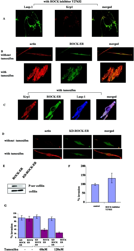
The transcription factor AP-1, which is composed of Fos and Jun family proteins, plays an essential role in tumor cell invasion by altering gene expression. We report here that Krp1, the AP-1 up-regulated protein that has a role in pseudopodial elongation in v-Fos-transformed rat fibroblast cells, forms a novel interaction with the nondifferentially expressed actin binding protein Lasp-1. Krp1 and Lasp-1 colocalize with actin at the tips of pseudopodia, and this localization is maintained by continued AP-1 mediated down-regulation of fibronectin that in turn suppresses integrin and Rho-ROCK signaling and allows pseudopodial protrusion and mesenchyme-like invasion. Mutation analysis of Lasp-1 demonstrates that its SH3 domain is necessary for pseudopodial extension and invasion. The results support the concept of an AP-1-regulated multigenic invasion program in which proteins encoded by differentially expressed genes direct the function, localization, and activity of proteins that are not differentially expressed to enhance the invasiveness of cells.


Figures

FIG. 1.
FIG. 1.
Krp1 can interact with nebulin repeat structures present in nebulin and Lasp-1. (A) Nebulin repeats of nebulin were subjected to in vitro transcription/translation, and GST pull-downs were performed with GST alone and GST-Krp1. (B) COS-7 cells were transfected with a Lasp-1-HA construct and the total cell lysate was used in a GST pull-down experiment with GST and GST-Krp1. Shown is a Coomassie-stained gel of purified GST and GST-Krp1 proteins and the Western blot of the pull-down which was probed with an HA antibody. (C) The reciprocal GST pull-down was performed by transfecting COS-7 cells with a Krp1-myc construct and the pull-down was performed with GST and GST Lasp-1. Shown is a Coomassie-stained gel of purified GST and GST-Lasp-1 proteins and the Western blot of the pull-down which was probed with a myc antibody. (D and E) FBR cells were transfected with a myc-tagged Krp1 construct (D) and with a Lasp-1-HA construct (E), and the cell lysates were used in coimmunoprecipitation with an anti-c-myc agarose (D) and anti-HA agarose (E). Shown is the Western blot analysis, which was probed with anti-Lasp-1 (D) and anti-Krp1 (E). IP, immunoprecipitation.
FIG. 2.
FIG. 2.
Localization of Lasp-1 in FBR and 208F cells. FBR (A) and 208F (B) cells were transiently transfected with a Lasp-1-GFP construct. (A) Confocal microscopy with Lasp-1-GFP and TRITC-phalloidin, to visualize F-actin. Boxed area shows the region that was enlarged to highlight the tips of the pseudopodia. (B) Confocal microscopy with Lasp-1-GFP and antipaxillin (red). Boxed area shows the region that was enlarged to highlight the localization of Lasp-1-GFP in focal adhesions and filopodia. (C) FBR cells transiently transfected with Lasp-1-GFP were incubated in a 3D invasion assay for 24 h; the figure shows the localization of Lasp-1-GFP after 24 h. Arrow highlights the accumulation of Lasp-1-GFP at the tips of pseudopodia.
FIG. 3.
FIG. 3.
Krp1 and Lasp-1 colocalize at the actin-rich membrane rufflelike structures at the tips of pseudopodia. (A) FBR cells were transiently transfected with Lasp-1-GFP. Shown are the confocal microscopy images with Lasp-1-GFP and anti-Krp1 (red). The boxed area shows the region that was enlarged to highlight the tips of pseudopodia. (B) Cells were permeablized in 0.02% Triton X-100 for 1 min and then fixed. (B) Confocal microscopy with anti-Krp1 (Green), TRITC-phalloidin, and anti-Lasp-1 (blue). Arrows highlight the tips of pseudopodia.
FIG. 4.
FIG. 4.
Expression of the dominant-negative Lasp-1 construct results in truncated pseudopodia and a decrease in the invasiveness of FBR cells in a 3D invasion assay. (A) FBR cells were transfected with Lasp-1ΔSH3-GFP construct; shown are confocal microscopy images with TRITC-phalloidin, anti-Krp1 (blue), and Lasp-1-GFP (green). The boxed region shows the area that was enlarged. (B) Quantitative analysis showing the percentage of transiently transfected cells that show a reduction in pseudopodium length. The experiment was repeated on three occasions. (C) FBR cells were transiently transfected with the following constructs: GFP, Krp1, Lasp-1, and Lasp-1ΔSH3 domain. The cells were allowed to invade in an in vitro 3D invasion assay for 3 days. Shown in the bar chart is the percentage of cells that have invaded. The experiment was repeated on three separate occasions.
FIG. 5.
FIG. 5.
siRNAs to Krp1 and Lasp-1 result in truncated pseudopodia. (A) Western blot analysis of FBR cells subjected to si control, siKrp1, and siLasp-1. (B) Quantitative analysis of the number of cells showing short pseudopodia when transfected with siKrp1, siLasp-1, and si control. (C) Cells were transfected with siKrp1, siLasp-1, and si control, and confocal microscopy was performed with TRITC-phalloidin, anti-Lasp-1 (green), and anti-Krp1 (blue).
FIG. 6.
FIG. 6.
Plating FBR cells on FN results in a loss of pseudopodium formation and the dissociation of Krp1 and Lasp-1. In addition, the presence of FN in a 3D invasion assay reduces the invasiveness of the cells. (A) FBR cells were plated on FN for 3 days; shown are confocal microscopy images with antipaxillin (green) and TRITC-phalloidin. (B) Confocal microscopy images of FBR cells grown on uncoated or FN-coated coverslips for 2 days and then transiently transfected with YFP-FAK. (C) FBR cells were plated on FN for 3 days, and shown are the confocal microscopy images of anti-Lasp-1 (green) and anti-Krp1 (red). The boxed region represents the cell which was enlarged. (D) Western blot analyses of FBR cells grown on uncoated and FN-coated coverslips that were subjected to treatment with 0.02% Triton X-100 before lysis. The Western blots were probed with anti-Krp1, anti-Lasp-1, and anti-extracellular regulated kinase 2 (anti-ERK2) as a loading control. (E) In vitro 3D invasion assay was performed with FBR cells with and without FN in the matrigel. Shown are the percentages of cells that invaded with and without FN in the matrigel. −Fn, without fibronectin, +Fn, with fibronectin.
FIG. 7.
FIG. 7.
RGD-containing inhibitory peptide blocks FBR cells spreading and stress fiber formation on FN. (A) FBR cells were plated onto FN-coated coverslips for 40 min in the presence of the inhibitory peptide (GRGDS) and the noninhibitory peptide (GRADSP); shown are the confocal microscopy images of antipaxillin (green) and TRITC-phalloidin (red). (B) FBR cells were plated onto FN-coated coverslips for 3 days and then subjected to either the GRGDS or the GRADSP peptides for 2 h; shown are the confocal microscopy images with antipaxillin (green) and TRITC-phalloidin (red). The boxed region represents the enlarged area.
FIG. 8.
FIG. 8.
The association of Krp1 and Lasp-1 and pseudopodium formation are Rho independent. FBR cells were grown on uncoated coverslips (A, B1, C, D, and E) and on FN-coated coverslips (B2). (A) Cells were transiently transfected with dominant-negative Rho (RhoN19-myc); shown are the confocal microscopy images with anti-myc (green) and TRITC-phalloidin (red). (B1 and 2) Cells were subjected to the Rho inhibitor C3 for 12 h; shown are the confocal microscopy images with anti-Krp1 (blue), anti-Lasp-1 (green), and TRITC-phalloidin. Cells grown on FN-coated coverslips without Rho inhibitor are shown in Fig. 6A and C. (C) Cells were transiently transfected with active Rho (RhoV14-myc) and shown are the confocal microscopy images with anti-myc (green) and TRITC-phalloidin. The boxed region represents the cell which was enlarged. Only the merged image is shown. (D) Cells were transiently transfected with active Rho (RhoV14-myc) and YFP-FAK; confocal microscopy images are shown with anti-myc (blue) and YFP-FAK (yellow). Only the merged image is shown. (E) Cells were transiently transfected with active Rho (RhoV14-myc) and Lasp-1-GFP; shown are the confocal microscopy images of anti-myc (blue), Lasp-1-GFP (green), and anti-Krp1 (red). The boxed region shows the cell that was enlarged. (F) GST-rhotekin pull-down assays were performed with lysates from FBR cells plated on uncoated and FN-coated tissue culture plates. Also shown is the input of Rho from each assay. Western blots were probed with anti-Rho A. (G and H) Western blot analysis of FBR cell lysates isolated from plating cells on FN and uncoated tissue culture plates. Western blots were probed with antibodies to ROCK 1 and 2 (G) and phospho-Ser cofilin (H). (I) FBR cells were grown on FN and uncoated coverslips with and without 10 μM of the ROCK inhibitor Y27632. Lysates were used for Western blotting; shown is the Western blot probed with anticofilin and anti-phospho-Ser cofilin.
FIG. 9.
FIG. 9.
Pseudopodium formation and invasion of FBR cells is ROCK independent. (A) FBR cells were grown on FN-coated coverslips for 3 days and then treated with the ROCK inhibitor Y27632 for 6 h; shown are the confocal microscopy images with anti-Lasp-1 (green) and anti-Krp1 (red). Cells grown on FN-coated coverslips without ROCK inhibitor are shown in Fig. 6A and C. Stable FBR cell lines expressing ROCK-ER-GFP (B and C) and kinase-dead ROCK-ER-GFP (D) were either treated with 4-hydroxytamoxifen for 24 h or were untreated. (B and D) Shown are the confocal microscopy images with and without tamoxifen with TRITC-phalloidin and ROCK-ER-GFP. (C) Confocal microscopy images with anti-Krp1 (red), ROCK-ER-GFP, and anti-Lasp-1 (blue). (E) Western blot analyses of cell lysates of stable FBR cell lines expressing ROCK-ER and kinase-dead ROCK-ER cells after tamoxifen treatment. Western blots were probed with anticofilin and anti-phospho-Ser cofilin. (F) In vitro 3D invasion assay was performed in the presence of ROCK inhibitor Y27632. The bar graph shows the percentages of cells that invaded in the absence and presence of Y27632. (G) In vitro 3D invasion assays were performed with stable FBR cells lines expressing ROCK-ER and kinase-dead ROCK-ER constructs. Shown are the percentages of cells that invaded after the induction of the constructs with various concentrations of tamoxifen.
FIG. 10.
FIG. 10.
Model showing how AP-1 mediates a multigenic invasion program.

Similar articles

Cited by

References

    1. Adams, J., R. Kelso, and L. Cooley. 2000. The kelch repeat superfamily of proteins: propellers of cell function. Trends Cell Biol. 10:17-24. - PubMed
    1. Akamatsu, H., K. Ichihara-Tanaka, K. Ozono, W. Kamiike, H. Matsuda, and K. Sekiguchi. 1996. Suppression of transformed phenotypes of human fibrosarcoma cells by overexpression of recombinant fibronectin. Cancer Res. 56:4541-4546. - PubMed
    1. Ali, I. U., V. Mautner, R. Lanza, and R. O. Hynes. 1977. Restoration of normal morphology, adhesion and cytoskeleton in transformed cells by addition of a transformation-sensitive surface protein. Cell 11:115-126. - PubMed
    1. Bahassi, E. M., S. Karyala, C. R. Tomlinson, M. A. Sartor, M. Medvedovic, and R. F. Hennigan. 2004. Critical regulation of genes for tumor cell migration by AP-1. Clin. Exp. Metastasis 21:293-304. - PubMed
    1. Bakin, A. V., and T. Curran. 1999. Role of DNA 5-methylcytosine transferase in cell transformation by fos. Science 283:387-390. - PubMed

Publication types

MeSH terms

LinkOut - more resources