. 2006 Feb;26(4):1510-7.
doi: 10.1128/MCB.26.4.1510-1517.2006.

Mouse disabled 1 regulates the nuclear position of neurons in a Drosophila eye model

Affiliations

Mouse disabled 1 regulates the nuclear position of neurons in a Drosophila eye model

Albéna Pramatarova et al. Mol Cell Biol. 2006 Feb.

Abstract

Nucleokinesis has recently been suggested as a critical regulator of neuronal migration. Here we show that Disabled 1 (Dab1), which is required for neuronal positioning in mammals, regulates the nuclear position of postmitotic neurons in a phosphorylation-site dependent manner. Dab1 expression in the Drosophila visual system partially rescues nuclear position defects caused by a mutation in the Dynactin subunit Glued. Furthermore, we observed that a loss-of-function allele of amyloid precursor protein (APP)-like, a kinesin cargo receptor, enhanced the severity of a Dab1 overexpression phenotype characterized by misplaced nuclei in the adult retina. In mammalian neurons, overexpression of APP reduced the ability of Reelin to induce Dab1 tyrosine phosphorylation, suggesting an antagonistic relationship between APP family members and Dab1 function. This is the first evidence that signaling which regulates Dab1 tyrosine phosphorylation determines nuclear positioning through Dab1-mediated influences on microtubule motor proteins in a subset of neurons.


Figures

FIG. 1.
FIG. 1.
Expression of tyrosine-phosphorylated Dab1RFP partially rescues the Gl1 external eye phenotype. (A and E) Scanning electron micrograph of an Elav-Gal4/+; +/+; Gl1/+ eye shows the characteristic small rough eye. At higher magnification the ommatidia appear disorganized, with variable sizes and frequent fusions. The bristles are also often duplicated. Bar, 100 μm. Panels E to H are magnified ×5 compared to panel A. (B and F) Gl1 flies overexpressing phosphorylated Dab1RFP (UAS-Dab1RFP/Elav-Gal4; +/+; Gl1/+) have fuller and smoother eyes, with a net improvement in the arrangement of ommatidial facets, fewer fusions, and better organized bristles. (C, D, G, and H) Flies expressing a phosphorylation-defective Dab1RFP-5F mutant (Elav-Gal4/+; UAS-Dab1RFP-5F/+; Gl1/+) (C and G) or RFP (UAS-RFP/Elav-Gal4; +/+; Gl1/+) (D and H) on the Gl1 background show no significant improvement of the external eye morphology. (I) The number of fusions in a 156- by 118-μm area of the eye was counted. Five or six animals were examined for each mutant. The data were analyzed by unpaired t tests (*, P < 0.0001; ♦, P = 0.1525; •, P = 0.6807). The error bars are standard errors of the means.
FIG. 2.
FIG. 2.
Photoreceptor cell differentiation and axonal projection in third-instar imaginal disks. (A) A wild-type third-instar eye disk stained with a photoreceptor-specific antibody (anti-Chaoptin, green), a neuronal nuclear marker (anti-Elav, blue) and phalloidin (red) demonstrates the stereotypical arrangement of photoreceptor clusters in the developing retina. The photoreceptor cell bodies are anchored in the upper two-thirds of the retina, between the actin-rich apical and basal surfaces. The optic stalk is free of nuclei. (B and C) In the Elav/+; +/+; Gl1/+ (B) or UAS-RFP/Elav; +/+; Gl1/+ (C) eye disks, the photoreceptors span the retina but many of the cell bodies and nuclei have migrated into the optic stalk. (D) However, in the UAS-Dab1RFP/Elav; +/+; Gl1/+ imaginal disks, fewer nuclei were observed in the lower one-third of the elongated retinal cells. Dab1RFP expression was observed throughout the cytoplasm of UAS-Dab1RFP/Elav;+/+;Gl1/+ photoreceptor cells (labeled with anti-Dab1 in red, phalloidin in green, and anti-Elav in blue). Bar, 10 μm.
FIG. 3.
FIG. 3.
Expression of phosphorylated Dab1RFP rescues the nuclear misplacement defect in the Gl1 mutant third-instar eye disk. (A) Wild-type photoreceptor nuclei (labeled with anti-Elav, green) are positioned in clusters under the actin-rich apical surface of the developing retina (red). Note that all nuclei are found in the top two-thirds of the retina and none in the optic stalk (bottom). Bar, 10 μm. (B) Schematic representation of a wild-type eye disk showing the arrangement of photoreceptor cells. MF, morphogenetic furrow; OS, optic stalk. (C) In the Gl1 animal the nuclei are scattered throughout the retina, and many of them migrate in the optic stalk, under the actin-rich basal surface of the retina. (D) Expression of phosphorylated Dab1RFP in the photoreceptor cells under the Elav-Gal4 driver (see Fig. 2) results in fewer nuclei trapped in the lower one-third of the retina and more found in their normal position closer to the apical surface. (E and F) Neither the phosphorylation-defective Dab1RFP-5F mutant (E) nor RFP alone (F) rescued the Gl1 nuclear phenotype. (G) The positions of individual nuclei were measured in four to six animals for each mutant (average of 300 to 500 nuclei per mutant). For each nucleus, the values were presented as a ratio of the total distance between the basal and apical surfaces over the distance of the individual nucleus to the apex. The values were grouped in two bins, with the basal bin (values of 1.5 or less) representing nuclei found in the lower one-third of the retina. The data were analyzed by unpaired t tests (*, P = 0.0353; ⧫, P = 0.3673; •, P = 0.6201). The error bars are standard errors of the means. WT, wild type.
FIG. 4.
FIG. 4.
Loss of APPL enhances the rough eye phenotype in Dab1RFP-overexpressing flies. (A and E) The scanning electron micrograph of an adult UAS-RFP/GMR-Gal4 control fly shows smooth external eye morphology with a linear array of ommatidial lens facets and interommatidial bristles. Bars, 100 μm (A) and 20 μm (E).(B and F) The UAS-Dab1RFP/GMR-Gal4 eye is rough, with disruptions in the linear arrangement of ommatidial lens facets and occasional fusions. (C and G) On the Appl-deficient background, UAS-RFP/GMR-Gal4 adults do not demonstrate any eye roughening. (D and H) However, expression of UAS-Dab1-RFP/GMR-Gal4 on the Appl-deficient background leads to worsening of the rough eye compared to that in panel B. The eye is characterized by a circular arrangement of the ommatidial lens facets compared to the relatively linear pattern observed in the UAS-Dab1-RFP/GMR-Gal4 flies, as well as more frequent ommatidial fusions.
FIG. 5.
FIG. 5.
Phosphorylated Dab1RFP alters nuclear or cellular positioning in the adult retina. (A) DAPI staining of control UAS-RFP/GMR-Gal4 eyes shows nuclei forming an arc pattern containing apical and basal nuclei with a nucleus-free region in the middle. Bar, 100 μm. (B) Adult UAS-Dab1RFP/GMR-Gal4 retinas had alterations in the nuclear position of the apical nuclei. (C) Minor disturbances of the outer layer of nuclei were observed in UAS-Dab1RFP-5F/GMR-Gal4 flies. (D) The arrangement of nuclei in UAS-Dab1RFP-158V/GMR-Gal4 flies was similar in severity to the Dab1RFP phenotype. (E) Appl deficiency causes only subtle, localized disturbances of the nuclear rows in Drosophila expressing RFP. (F) However, Appl deficiency in conjunction with the UAS-Dab1RFP/GMR-Gal4 transgenes caused a dramatic increase in the number of misplaced nuclei, with a loss of the central nucleus-free area. (G) The nuclear placement in Dab1RFP-5F-expressing retinas was also more erratic on the Appl mutant background. (H) In contrast, the phenotype of Dab1RFP-158V/GMR-Gal4 was relatively unaltered by Appl deficiency.
FIG. 6.
FIG. 6.
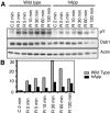
APP transgenic neurons respond less robustly to Reelin stimulation. (A) Primary neuronal cultures from embryonic day 15.5 wild-type or hAPP transgenic littermates were stimulated with control (C) or Reelin-enriched (R) medium for 2 to 120 min as indicated. Reelin-induced Dab1 tyrosine phosphorylation (pY) was apparent after 2 min of treatment, with a peak at 30 min, followed by signal decrease over the next 1.5 h in wild-type neurons. In contrast, stimulation of hAPP-expressing neurons failed to induce equivalent levels of Dab1 tyrosine phosphorylation. Similar time courses and total phosphorylation levels were observed in five paired experiments. (B) Quantification of Dab1 tyrosine phosphorylation normalized to actin from the experiment shown in panel A demonstrated that hAPP expression reduces Dab1 tyrosine phosphorylation to a value of less than half of that observed from wild-type neurons.
FIG. 7.
FIG. 7.
A model for the role of Dab1 in nuclear placement. Enhancement of dynein-dynactin-related activity and suppression of Kinesin function are possible mechanisms by which tyrosine-phosphorylated Dab1 could act to partially rescue the Glued mutant phenotype.

Similar articles

Cited by

References

    1. Arnaud, L., B. A. Ballif, and J. A. Cooper. 2003. Regulation of protein tyrosine kinase signaling by substrate degradation during brain development. Mol. Cell. Biol. 23:9293-9302. - PMC - PubMed
    1. Arnaud, L., B. A. Ballif, E. Förster, and J. A. Cooper. 2003. Fyn tyrosine kinase is a critical regulator of Disabled-1 during brain development. Curr. Biol. 13:9-17. - PubMed
    1. Assadi, A. H., G. Zhang, U. Beffert, R. S. McNeil, A. L. Renfro, S. Niu, C. C. Quattrocchi, B. A. Antalffy, M. Sheldon, D. D. Armstrong, A. Wynshaw-Boris, J. Herz, G. D'Arcangelo, and G. D. Clark. 2003. Interaction of reelin signaling and Lis1 in brain development. Nat. Genet. 35:270-276. - PubMed
    1. Ballif, B. A., L. Arnaud, W. T. Arthur, D. Guris, A. Imamoto, and J. A. Cooper. 2004. Activation of a Dab1/CrkL/C3G/Rap1 pathway in Reelin-stimulated neurons. Curr. Biol. 14:606-610. - PubMed
    1. Bock, H. H., and J. Herz. 2003. Reelin activates SRC family tyrosine kinases in neurons. Curr. Biol. 13:18-26. - PubMed

Publication types

MeSH terms

LinkOut - more resources