. 2006 Feb;26(4):1518-26.
doi: 10.1128/MCB.26.4.1518-1526.2006.

The NH2 tail of the novel histone variant H2BFWT exhibits properties distinct from conventional H2B with respect to the assembly of mitotic chromosomes

Affiliations

The NH2 tail of the novel histone variant H2BFWT exhibits properties distinct from conventional H2B with respect to the assembly of mitotic chromosomes

Mathieu Boulard et al. Mol Cell Biol. 2006 Feb.

Abstract

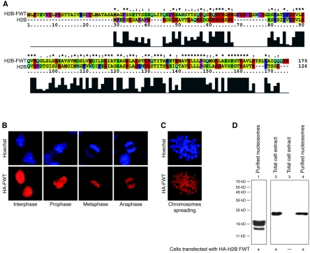
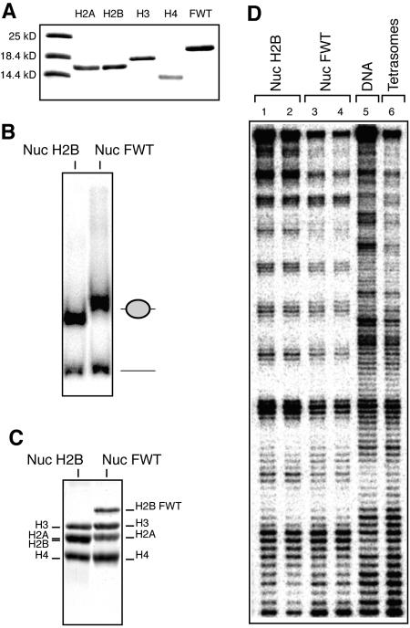
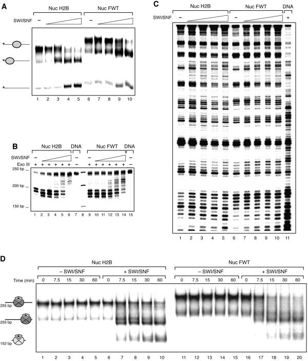
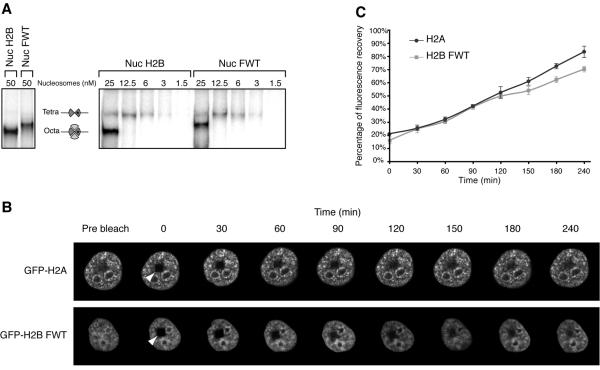
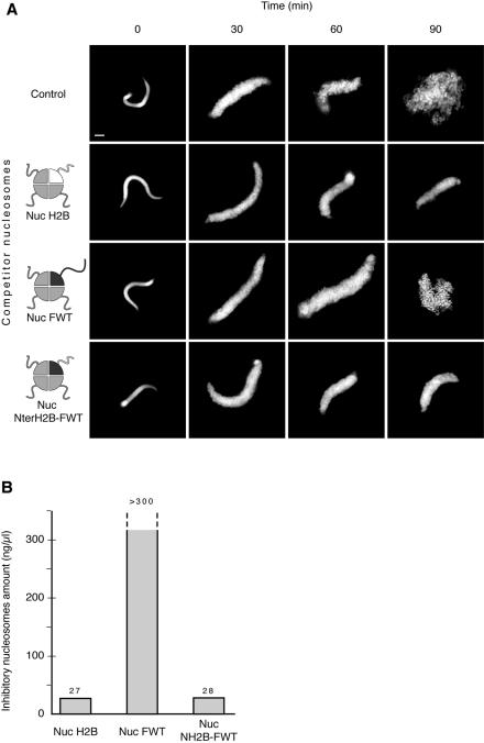
We have studied the functional and structural properties of nucleosomes reconstituted with H2BFWT, a recently identified putative histone variant of the H2B family with totally unknown function. We show that H2BFWT can replace the conventional histone H2B in the nucleosome. The presence of H2BFWT did not affect the overall structure of the nucleosome, and the H2BFWT nucleosomes exhibited the same stability as conventional nucleosomes. SWI/SNF was able to efficiently remodel and mobilize the H2BFWT nucleosomes. Importantly, H2BFWT, in contrast to conventional H2B, was unable to recruit chromosome condensation factors and to participate in the assembly of mitotic chromosomes. This was determined by the highly divergent (compared to conventional H2B) NH2 tail of H2BFWT. These data, in combination with the observations that H2BFWT was found by others in the sperm nuclei and appeared to be associated with the telomeric chromatin, suggest that H2BFWT could act as a specific epigenetic marker.


Figures

FIG. 1.
FIG. 1.
The histone variant H2BFWT is assembled into nucleosomes. (A) Sequence alignment of conventional H2B and the histone variant H2BFWT. The histograms under the sequences show the degree of homology between the two proteins. The amino acid residues are shown in different colors according to their properties. (B and C) In vivo association of H2BFWT with chromatin. HeLa cells were transiently transfected with a tagged HA-H2BFWT expression vector. The presence of the tagged HA-H2BFWT at different phases of the cell cycle (B) and in spread metaphase chromosomes (C) was detected with anti-HA antibody (red). Hoechst staining of DNA is shown in blue. (D) Incorporation of H2BFWT into the nucleosomes. Nuclei isolated from HA-H2BFWT-transfected cells were digested with microccocal nuclease, and nucleosomes were purified on a sucrose gradient containing 0.6 M NaCl. The presence of H2BFWT was detected by Western blotting by using anti-HA antibody. Lane 1, Coomassie blue staining of an 18% sodium dodecyl sulfate gel of the loaded nucleosomes. Lanes 2 to 4, Western blotting of extracts isolated from H2BFWT-transfected cells (lane 2), control nontransfected cells (lane 3), and isolated nucleosomes (lane 4). The positions of the molecular mass markers are shown on the left.
FIG. 2.
FIG. 2.
The presence of H2BFWT has no effect on the structure of the nucleosome. An equimolar mixture of purified-to-homogeneity recombinant core histones H2A, H3, and H4 and either conventional H2A or histone variant H2BFWT and a 152-bp EcoRI-RsaI 32P-end-labeled DNA fragment containing the X. borealis 5S RNA gene were used to reconstitute nucleosomes. (A) An 18% sodium dodecyl sulfate electrophoresis of conventional core histones and the histone variant H2BFWT. The first lane shows the molecular mass markers. (B) EMSA of the reconstituted conventional (Nuc) and histone variant H2BFWT (Nuc FWT) nucleosomes. The positions of the nucleosomes and the free DNA are shown on the right. (C) Histone composition of the reconstituted nucleosomes. Preparative EMSA was carried out, the conventional and H2BFWT nucleosome bands were cut from the gel, and the nucleoproteins were eluted. Histones were isolated and separated by 18% sodium dodecyl sulfate electrophoresis, and the gel was stained with Coomassie blue. The positions of the histones are indicated. (D) DNase I footprinting of reconstituted conventional H2AB nucleosomes (in duplicate, lanes 1 and 2), variant H2AB nucleosomes (in duplicate, lanes 3 and 4), and tetrasomal particles containing only the (H3-H4)2 tetramer (lane 6). Lane 5, footprinting of naked DNA.
FIG. 3.
FIG. 3.
SWI/SNF remodels and mobilizes conventional and variant H2BFWT nucleosomes. (A) A 255-bp DNA fragment containing the positioning sequence 601 was used to reconstitute conventional and variant H2BFWT centrally positioned nucleosomes. Both types of nucleosomes were incubated with increasing amounts of SWI/SNF in the presence of ATP for 30 min at 30°C. The mobility of the histone octamers was assessed on a 5.5% native polyacrylamide gel. On the left are indicated the positions of the centrally and end-positioned nucleosomes and free DNA. (B) Exonuclease III mapping of the positions of conventional and H2BFWT nucleosomes. Both types of nucleosomes were incubated with SWI/SNF as described for panel A and, after the reaction was arrested, digested with exonuclease III. (C) DNase I footprinting of conventional (lanes 1 to 5) and variant H2BFWT (lanes 6 to 10) nucleosomes, reconstituted on the 152-bp EcoRI-RsaI 32P-end-labeled DNA fragment containing the X. borealis 5S RNA. The nucleosomes were incubated with SWI/SNF as for panel A and then subjected to DNase I digestion. Lane 11, DNase I digestion pattern of naked DNA. (D) The H2A-H2BFWT dimer is transferred efficiently to an (H3-H4)2 tetrameric particle. Conventional and H2BFWT variant nucleosomes were reconstituted on a 255-bp DNA fragment which contains the 601 positioning sequence by using radioactively labeled H2A. Histone H3-H4 particles were reconstituted on a 152-bp fragment containing the X. borealis 5S gene. The nucleosomes were incubated in the presence of a threefold molar excess of tetrameric particles at 30°C for the indicated times in the presence or absence of SWI/SNF. The positions of the centrally and end-positioned 255-bp nucleosomes and of the 152-bp nucleosomes are indicated. The asterisk indicates the radioactively labeled H2A. Note that the conventional H2A-H2B and the variant H2A-H2BFWT dimers exhibit very similar efficiencies of transfer to the tetrameric particles.
FIG. 4.
FIG. 4.
In vitro and in vivo stabilities of the variant H2BFWT nucleosome. (A) The H2BFWT variant nucleosome exhibits in vitro the same stability as the conventional H2A nucleosome. Conventional and variant H2BFWT nucleosomes and tetrameric (H3-H4)2 particles were reconstituted by using 32P-labeled histone H3. The samples were diluted to the indicated concentrations with 1× phosphate-buffered saline and run on a native 5.5% polyacrylamide gel. On the left are shown the conventional and H2BFWT nucleosomes at 50 nM. The positions of the intact nucleosomes and tetrameric particles are indicated. Note that upon dilution, the conventional H2A-H2B and the variant H2A-H2BFWT dimers are released with essentially the same efficiency from the reconstituted nucleosomes. (B) FRAP analysis of the mobility of the GFP-H2BFWT variant fusion. Cells stably expressing either GFP-H2A or GFP-H2BFWT were photobleached, and images were recorded at the indicated times postbleaching. The rectangular bleached area is designated by an arrowhead. Images of the cell before bleaching (prebleach) are also shown. (C) Quantification of the data shown in panel B. The means and standard deviations for 45 nuclei from five independent experiments are shown.
FIG. 5.
FIG. 5.
The assembly of mitotic chromosomes in Xenopus egg extract is not inhibited by the variant H2BFWT nucleosomes, in contrast to conventional nucleosomes. (A) Demembranated sperm nuclei were incubated in the extract for the times indicated in the absence (control) or in the presence of either conventional H2B (Nuc H2B), variant H2BFWT (Nuc FWT), or fusion NterH2B-histone fold H2BFWT (Nuc NterH2B-FWT) nucleosomes at a concentration of 30 ng per μl of extract. After fixation and staining with Hoechst 33258, the assembled structures were observed by fluorescence microscopy. The reconstituted particles are schematically presented on the left. Conventional H2B is in white, while H2BFWT is in black. Note that both the conventional and the fusion NterH2B-histone fold H2BFWT nucleosomes inhibit chromosome assembly, while the variant H2BFWT nucleosomes do not. Bar, 5 μm. (B) Quantification of the minimal nucleosome amount that is able to inhibit mitotic chromosome condensation. Increasing amount of conventional H2B (Nuc H2B), variant H2BFWT (Nuc FWT), or fusion NterH2B-histone fold H2BFWT (Nuc NterH2B-FWT) nucleosomes were added to the chromosome assembly reaction mixture, and the minimal concentration of nucleosomes sufficient to inhibit chromosome condensation was measured. Note that even the use of 300 ng/μl of competitor H2BFWT nucleosomes (the highest nucleosomal concentration possible to be used in the experiments) was unable to inhibit the assembly process.

Similar articles

Cited by

References

    1. Allan, J., N. Harborne, D. C. Rau, and H. Gould. 1982. Participation of core histone “tails” in the stabilization of the chromatin solenoid. J. Cell Biol. 93:285-297. - PMC - PubMed
    1. An, W., V. B. Palhan, M. A. Karymov, S. H. Leuba, and R. G. Roeder. 2002. Selective requirements for histone H3 and H4 N termini in p300-dependent transcriptional activation from chromatin. Mol. Cell 9:811-821. - PubMed
    1. Angelov, D., A. Molla, P. Y. Perche, F. Hans, J. Cote, S. Khochbin, P. Bouvet, and S. Dimitrov. 2003. The Histone variant macroH2A interferes with transcription factor binding and SWI/SNF nucleosome remodeling. Mol. Cell 11:1033-1041. - PubMed
    1. Angelov, D., A. Verdel, W. An, V. Bondarenko, F. Hans, C. M. Doyen, V. M. Studitsky, A. Hamiche, R. G. Roeder, P. Bouvet, and S. Dimitrov. 2004. SWI/SNF remodeling and p300-dependent transcription of histone variant H2ABbd nucleosomal arrays. EMBO J. 23:3815-3824. - PMC - PubMed
    1. Arents, G., R. W. Burlingame, B.-C. Wang, W. E. Love, and E. N. Moudrianakis. 1991. The nucleosomal core histone octamer at 3.1 A resolution: a tripartite protein assembly and a left-handed superhelix. Proc. Natl. Acad. Sci. USA 88:10148-10152. - PMC - PubMed

Publication types

MeSH terms

LinkOut - more resources