. 2006 Feb 27;94(4):513-23.
doi: 10.1038/sj.bjc.6602971.

Inhibitory effects of retinoic acid metabolism blocking agents (RAMBAs) on the growth of human prostate cancer cells and LNCaP prostate tumour xenografts in SCID mice

Affiliations

Inhibitory effects of retinoic acid metabolism blocking agents (RAMBAs) on the growth of human prostate cancer cells and LNCaP prostate tumour xenografts in SCID mice

C K Huynh et al. Br J Cancer. .

Abstract

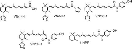
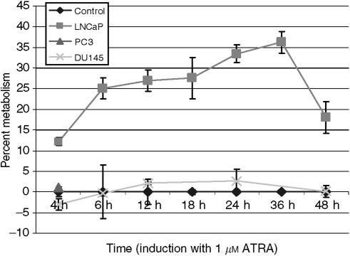
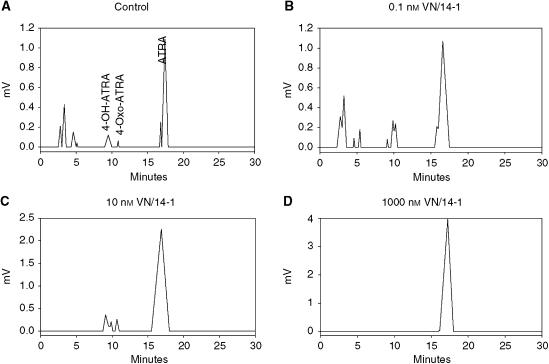
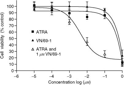
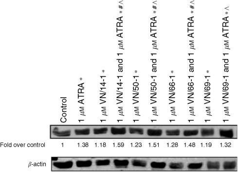
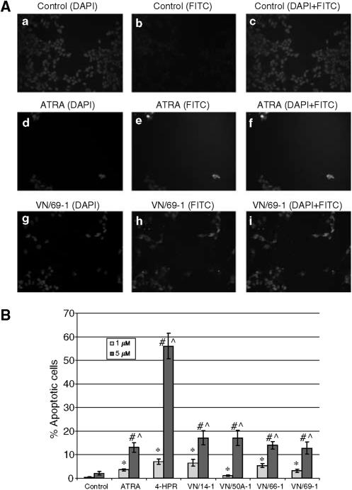
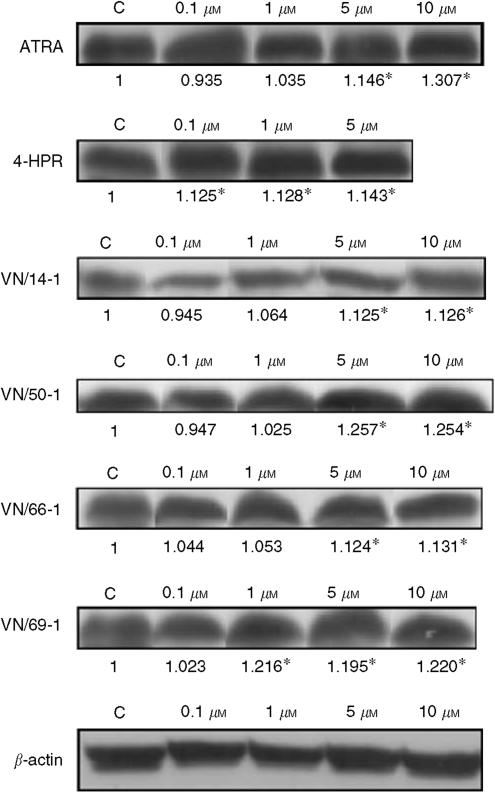
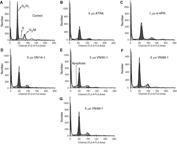
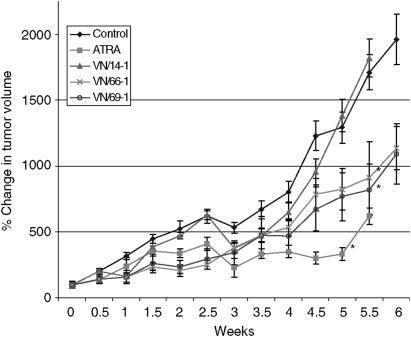
In recent studies, we have identified several highly potent all-trans-retinoic acid (ATRA) metabolism blocking agents (RAMBAs). On the basis of previous effects of liarozole (a first-generation RAMBA) on the catabolism of ATRA and on growth of rat Dunning R3227G prostate tumours, we assessed the effects of our novel RAMBAs on human prostate tumour (PCA) cell lines. We examined three different PCA cell lines to determine their capacity to induce P450-mediated oxidation of ATRA. Among the three different cell lines, enhanced catabolism was detected in LNCaP, whereas it was not found in PC-3 and DU-145. This catabolism was strongly inhibited by our RAMBAs, the most potent being VN/14-1, VN/50-1, VN/66-1, and VN/69-1 with IC50 values of 6.5, 90.0, 62.5, and 90.0 nM, respectively. The RAMBAs inhibited the growth of LNCaP cells with IC50 values in the microM-range. In LNCaP cell proliferation assays, VN/14-1, VN/50-1, VN/66-1, and VN/69-1 also enhanced by 47-, 60-, 70-, and 65-fold, respectively, the ATRA-mediated antiproliferative activity. We then examined the molecular mechanism underlying the growth inhibitory properties of ATRA alone and in combination with RAMBAs. The mechanism appeared to involve the induction of differentiation, cell-cycle arrest, and induction of apoptosis (TUNEL), involving increase in Bad expression and decrease in Bcl-2 expression. Treatment of LNCaP tumours growing in SCID mice with VN/66-1 and VN/69-1 resulted in modest but statistically significant tumour growth inhibition of 44 and 47%, respectively, while treatment with VN/14-1 was unexpectedly ineffective. These results suggest that some of our novel RAMBAs may be useful agents for the treatment of prostate cancer.


Figures

Figure 1
Figure 1
Chemical structures of RAMBAs, VN/14-1, VN/50-1, VN/66-1, and VN/69-1 and 4-HPR.
Figure 2
Figure 2
Time course of induction of CYP26 in prostate cancer cells. LNCaP, PC3, and DU145 cells were pre-incubated with 1 μM ATRA for various time periods to induce the CYP26 enzyme. Cells were then isolated and incubated at 37°C for 5 h with 0.8 μM [11,12-3H]-ATRA. Retinoids were extracted, and metabolites analysed by HPLC as described in Materials and Methods. Metabolic activity was calculated as percentage of polar ATRA metabolites of total radioactivity. The metabolic activity of induced LNCaP, PC3, and DU145 cells was then divided by the metabolic activity of the respective un-induced cells (percent metabolism). The percent metabolism of each un-induced cell line was set to the x-axis (Control) and the percent metabolism of the induced cell lines was plotted accordingly. The experiments were performed thrice.
Figure 3
Figure 3
Inhibition of ATRA metabolism by VN/14-1 in intact LNCaP cells. Human LNCaP prostate cancer cells were cultured under basal conditions (data not shown) or pretreated with 1 μM ATRA (AD). Thereafter, cells were washed, and incubated with 0.1 μM [11,12-3H]-ATRA, either in the absence (A) or presence (BD) of VN/14-1 at concentrations of 0.1, 10, and 1000 nM, respectively. The cells and media were collected, extracted and analysed by reverse phase HPLC as described in Materials and Methods. The experiments were performed twice.
Figure 4
Figure 4
Antiproliferative effects of ATRA or VN/69-1 alone and ATRA in combination with VN/69-1 (1 μM). LNCaP cells were incubated with various concentrations of ATRA, the RAMBAs alone and in combination. The MTT assay was performed. The plot represents the percentage of viable cells vs the concentration of agents used. The IC50 value was determined as the concentration of agents that inhibited the viability of LNCaP cells by 50%. Similar results were obtained for VN/14-1, VN/50-1, VN/66-1, and 4-HPR.
Figure 5
Figure 5
Effects of ATRA and RAMBAs alone and in combination on levels of cytokeratin 18 (a differentiation marker) in human prostate LNCaP cells. Cells were incubated with ATRA or RAMBAs alone or in combinations for 6 days. Lysates were subjected to SDS–PAGE and Western blotting. Membranes were probed with cytokeratin 18 antibody, and intensities of bands were analysed by densitometry. Groups labelled with * are significantly different from control (P<0.05). Groups labelled with # are significantly different from ATRA alone (P<0.05). Groups labelled with ^ are significantly different between combination RAMBA with ATRA compared to the corresponding RAMBA alone (P<0.05).
Figure 6
Figure 6
(A) Apoptosis in LNCaP cells determined by TUNEL and analysed by fluorescence microscopy. LNCaP cells were treated with 5 μM ATRA or RAMBA for 6 days. Cells were then fixed, stained, mounted, and examined under fluorescence microscopy. Control is shown in a, b, and c. ATRA (5 μM) is shown in d, e, and f. VN/69-1 (5 μM) is shown in g, h, and i. Nuclear DAPI staining is shown in a, d, and g. FITC staining for nicked DNA is shown in b, e, and h. Combined DAPI and FITC staining is shown in c, f, and i. (B) TUNEL analysis of LNCaP cells treated with ATRA, 4-HPR, or RAMBAs. LNCaP cells were incubated with either 1 or 5 μM of ATRA, 4-HPR, or a RAMBA for 6 days. LNCaP cells were then fixed, stained, mounted, and analysed by fluorescence microscopy. The number of apoptotic cells and total number of cells were counted in each of the five fields for each treatment and the percentage of apoptotic cells was calculated and plotted. Treatment with 1 μM drugs significantly increased from its control (*) (P<0.05). Treatment with 5 μM drugs significantly increased from its control (#) (P<0.05). Treatment with 5 μM of drugs significantly increased from 1 μM of the same drug (^) (P<0.05).
Figure 7
Figure 7
Western immunobloting of whole-cell lysates of treated LNCaP cells for expression of Bad. Cell lysates were used as described in the differentiation assay. Bad is a proapoptotic protein. The Western blots of Bad expressions are shown. The lanes are labelled above the blots and the expression of Bad is expressed as fold over control as determined by densitometry (below the blot). There was a significant difference in groups labelled with * compared to control (P<0.05).
Figure 8
Figure 8
Cell-cycle analysis of LNCaP cells treated with 1 or 5 μM ATRA, 4-HPR or RAMBAs. LNCaP cells were incubated with either 1 or 5 μM ATRA, 4-HPR, or RAMBA for 6 days. LNCaP cells were then fixed, stained with propidium iodide, and analysed by FACScan. Histograms of the FACScan analysis from control (A), ATRA (B), 4-HPR (C), VN/14-1 (D), VN/50A-1 (E), VN/66-1 (F), and VN/69-1 (G) are shown.
Figure 9
Figure 9
The effect of ATRA, VN/14-1, VN/66-1 on LNCaP tumour volumes in male SCID mice. Male SCID mice were inoculated s.c. with LNCaP cells suspended in Matrigel at two sites in the flank. The sizes of the tumours were determined by measuring the tumour volumes using calipers. Tumour volumes were calculated using the formula V=4/3 × π × r12 × r2 (r1<r2). The tumours were allowed to develop to 100 mm3 before treatment. The mice were treated with an equivalent dose to 0.033 mmol kg−1 of ATRA, VN/14-1, VN/66-1 or VN/69-1. The vehicle control was hydroxypropyl-β-cyclodextrin (HPC) in saline. (*) indicated that these treatment groups were significantly different from vehicle control (P<0.05).

Similar articles

Cited by

References

    1. Belosay A, Njar VCO, Brodie AMH (2005) Effects and molecular mechanisms of novel retinoic acid metabolism blocking agent (VN/14-1) in MCF-7Ca cells insensitive to aromatase inhibitor letrozole. Clin Cancer Res (in review) - PubMed
    1. Berges RS, Vukanovic J, Epstein JI, Carmichel M, Cisek L, Johnson DE, Veltri RW, Walsh PH, Isaacs JT (1995) Implications of cell kinetic changes during progression of human prostatic cancer. Clin Cancer Res 1: 475–481 - PMC - PubMed
    1. Bollag W, Isnardi L, Jablonski S, Klaus M, Majewski S, Pirson W, Toma S (1997) Links between pharmacological properties of retinoids and nuclear retinoid receptors. Int J Cancer 70: 470–472 - PubMed
    1. Campbell MJ, Park S, Uskokovic MR, Dowson MI, Koeffler HP (1998) Expression of retinoic acid receptor-beta sensitizes prostate cancer cells to growth inhibition mediated by combinations with retinoids and a 19-nor hexafluoride vitamin D3 analog. Endocrinology 139: 1927–1980 - PubMed
    1. Carter BS, Carter B, Issacs JT (1990) Epidemiological evidence regarding predisposing factors to prostate cancer. Prostate 16: 187–197 - PubMed

Publication types