. 2006 Mar;133(5):967-75.
doi: 10.1242/dev.02270. Epub 2006 Feb 1.

Transgenic isolation of skeletal muscle and kidney defects in laminin beta2 mutant mice: implications for Pierson syndrome

Affiliations

Transgenic isolation of skeletal muscle and kidney defects in laminin beta2 mutant mice: implications for Pierson syndrome

Jeffrey H Miner et al. Development. 2006 Mar.

Abstract

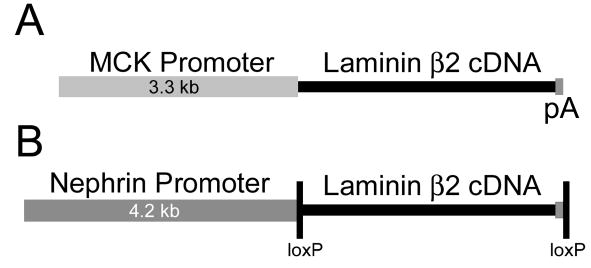
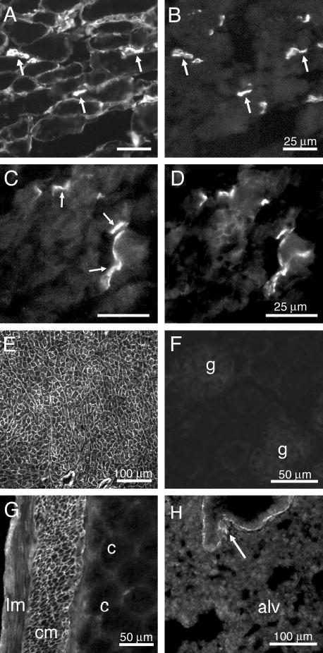
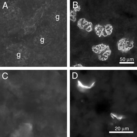
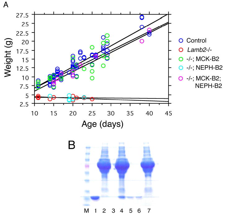
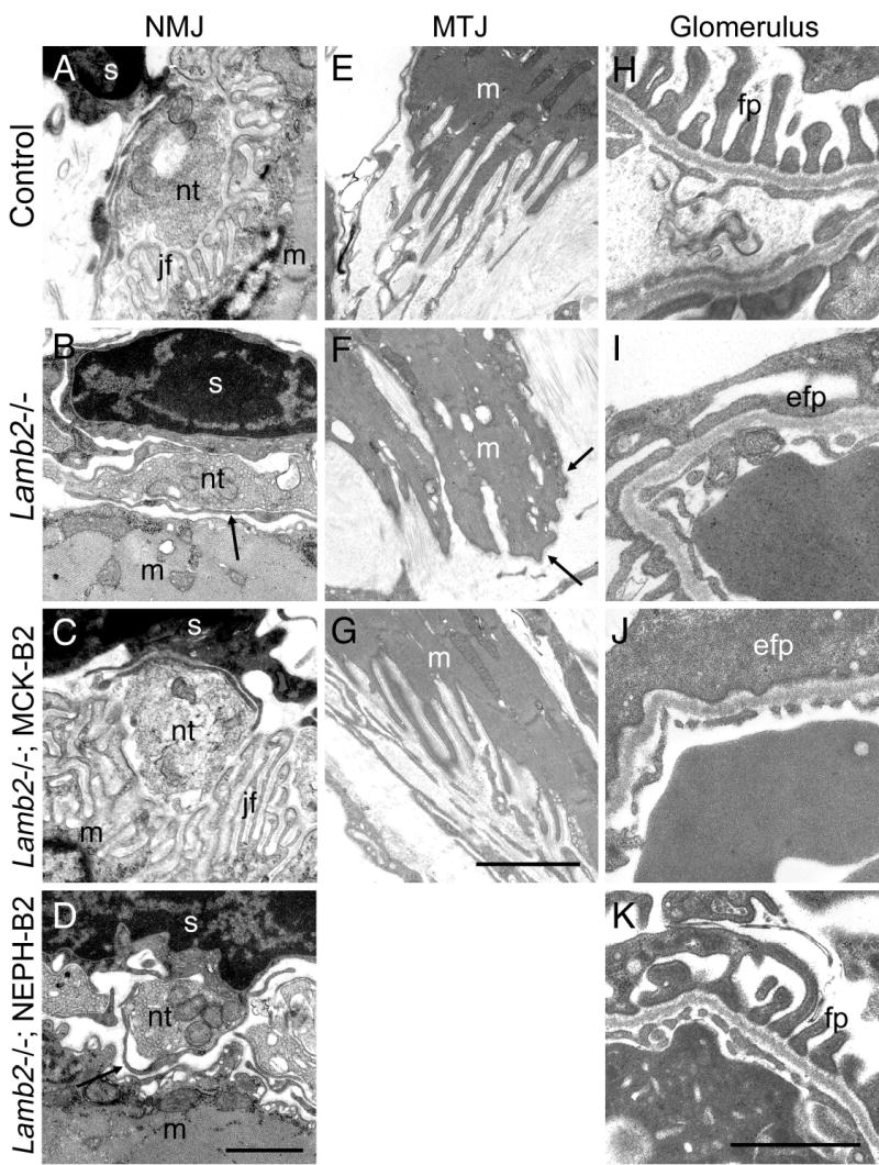
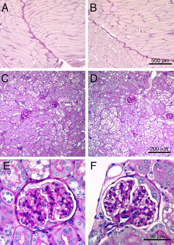
Pierson syndrome is a recently defined disease usually lethal within the first postnatal months and caused by mutations in the gene encoding laminin beta2 (LAMB2). The hallmarks of Pierson syndrome are congenital nephrotic syndrome accompanied by ocular abnormalities, including microcoria (small pupils), with muscular and neurological developmental defects also present. Lamb2(-/-) mice are a model for Pierson syndrome; they exhibit defects in the kidney glomerular barrier, in the development and organization of the neuromuscular junction, and in the retina. Lamb2(-/-) mice fail to thrive and die very small at 3 weeks of age, but to what extent the kidney and neuromuscular defects each contribute to this severe phenotype has been obscure, though highly relevant to understanding Pierson syndrome. To investigate this, we generated transgenic mouse lines expressing rat laminin beta2 either in muscle or in glomerular epithelial cells (podocytes) and crossed them onto the Lamb2(-/-) background. Rat beta2 was confined in skeletal muscle to synapses and myotendinous junctions, and in kidney to the glomerular basement membrane. In transgenic Lamb2(-/-) mice, beta2 deposition in only glomeruli prevented proteinuria but did not ameliorate the severe phenotype. By contrast, beta2 expression in only muscle restored synaptic architecture and led to greatly improved health, but the mice died from kidney disease at 1 month. Rescue of both glomeruli and synapses was associated with normal weight gain, fertility and lifespan. We conclude that muscle defects in Lamb2(-/-) mice are responsible for the severe failure to thrive phenotype, and that renal replacement therapy alone will be an inadequate treatment for Pierson syndrome.


Figures

Fig. 1
Fig. 1
Structure of the transgenes. (A) The MCK-B2 transgene drives rat laminin β2 expression from the mouse muscle creatine kinase (MCK) promoter, and transcription termination and polyadenylylation signal sequences from SV40 (pA) ensure processing to mRNA. (B) The NEPH-B2 transgene drives β2 expression from the mouse nephrin promoter. The β2 cDNA and SV40 sequences were flanked by loxP sites for future manipulations.
Fig. 2
Fig. 2
Localization of endogenous and MCK-B2 transgene-derived laminin β2. (A,B) In control skeletal muscle, endogenous mouse laminin β2 (A) is concentrated at synapses (arrows) doubly labeled by α-bungarotoxin (B); β2 is also found in extrasynaptic regions of muscle fibers (A). (C,D) In MCK-B2 transgenics, antibody specific for transgene-derived rat β2 (C) only labels synapses in skeletal muscle (arrows), identified by α-bungarotoxin (D). (E-H) Transgene-derived rat laminin β2 is also found in cardiac muscle BMs (E), in circular (cm) but not longitudinal smooth muscle (lm) or crypt (c) epithelial BMs of intestine (G), and weakly in large airway smooth muscle of lung (arrow in H) but not in alveolar (alv) BMs. No rat β2 was detected in glomeruli (g) in kidney (F). Scale bars in A-D, 25 μm; in E and H, 100 μm; in F and G, 50 μm.
Fig. 3
Fig. 3
NEPH-B2 transgene-derived laminin β2 accumulates specifically in the GBM. (A,B) Antibody specific for transgene-derived rat β2 does not stain kidney glomeruli (g) from a control mouse (A) but stains GBM in kidney from NEPH-B2 transgenic mice (B). (C,D) NEPH-B2 transgene-derived β2 is not deposited at skeletal muscle synapses (C) identified by double staining with α-bungarotoxin (D). Scale bar in B, 50 μm; in D, 20 μm.
Fig. 4
Fig. 4
(A) Growth curves (weight vs. age) of control mice, Lamb2−/− mice, and Lamb2−/− mice carrying transgenes. Lamb2−/− and Lamb2−/−; NEPH-B2 mice fail to surpass a weight of ~4 g and die at 3 to 4 weeks of age. In contrast, Lamb2−/−; MCK-B2 mice grow at a rate similar to controls but die at 1 month of age. Lamb2−/−; MCK-B2; NEPH-B2 doubly transgenic mice exhibit normal weight gain and a long life. (B) Proteinuria in Lamb2−/−; MCK-B2 mice. One μL of urine was analyzed by SDS-PAGE and stained with Coomassie blue. M, markers; the pink band is albumin. Lanes 1, 5, and 6: urine from Lamb2+/−; MCK-B2 mice. Lanes 2, 4, and 7: urine from Lamb2−/−; MCK-B2 mice. Lane 3 was empty.
Fig. 5
Fig. 5
Restoration of proper synaptic architecture and BM composition in skeletal muscle from Lamb2−/−; MCK-B2 mice. (A,B) Colocalization of rat laminin β2 (A) with α-bungarotoxin (B) in a highly ramified skeletal muscle synapse. (C,D) Restoration of laminin α5 to synaptic BMs (C) identified by α-bungarotoxin (D). (E,F) Laminin β1 (E, green in F) is not associated with the α-bungarotoxin-positive synaptic clefts (red in the merged image in F). Scale bar in F, 20 μm for A-F.
Fig. 6
Fig. 6
Ultrastructural analysis of neuromuscular junctions (NMJ), myotendinous junctions (MTJ), and glomerular filtration barriers. (A-D) A control synapse (A) shows a Schwann cell (s) capping the vesicle-rich nerve terminal (nt) adjacent to the muscle (m) endplate containing numerous junctional folds (jf). In the Lamb2−/− synapse (B), junctional folds are absent and the Schwann cell extends processes (arrow) between the nerve terminal and the muscle. Synaptic deposition of laminin β2 in Lamb2−/−; MCK-B2 mice restores synaptic architecture to normal (C). In Lamb2−/−; NEPH-B2 mice, glomerular deposition of β2 and prevention of proteinuria has no restorative effect on the synapse (D). (E-G) MTJ from a control (E) exhibits numerous infoldings of the muscle fiber (m) with continuous BMs. In the Lamb2−/− MTJ (F), infoldings are less complex, and the BMs (arrows) appear fuzzy. MCK-B2 transgene-derived β2 restores much of the normal MTJ architecture (G). (H-K) Glomerular capillary segment from a control (H) shows the interdigitated podocyte foot processes (fp) adjacent to the GBM. Effaced foot processes (efp) are evident in the Lamb2−/− (I) and Lamb2−/−; MCK-B2 (J) mice, which are proteinuric. Deposition of rat β2 into the GBM in Lamb2−/−; NEPH-B2 mice prevents proteinuria and foot process effacement (K). Scale bars in D and K are 1 μm for A-D and H-K, respectively; scale bar in G is 2 μm for E-G.
Fig. 7
Fig. 7
Functional and compositional analyses of MTJs. (A-D) Evans blue analysis reveals normal MTJ integrity in a control mouse diaphragm (A), but evidence of damage is observed in Lamb2−/− (B) and Lamb2−/−; MCK-B2 (C) mice at 3 weeks of age. A diaphragm from an older adult Lamb2−/−; MCK-B2; NEPH-B2 mouse shows no damage (D), suggesting eventual repair of MTJs. (E,F) Double staining for laminin β2 (green) and laminin α2 (red) in intercostal muscles reveals concentration of β2 at MTJs in both control (E) and Lamb2−/−; MCK-B2 (F) mice. (G,H) Double staining for mouse plus rat β2 (G) and rat β2 only (H) in a Lamb2+/−; MCK-B2 mouse shows that many MTJs contain little if any transgene-derived protein. (I-L) Double staining for total laminin β2 (green) and α2 (red) in intercostal muscles from aged control (I,J) and Lamb2−/−; MCK-B2; NEPH-B2 (K,L) mice reveals efficient accumulation of β2 at MTJs. Note the lack of transgene-derived β2 in extra-junctional BMs in rescued mutant muscle fibers (F,K). Scale bar in D, 1 mm for A-D; in H, 50 μm for A-I and K; in L, 20 μm for J and L.
Fig. 8
Fig. 8
Analysis of laminin β chain composition in glomeruli. (A,B) In Lamb2+/−; NEPH-B2 mice, the GBM contains rat laminin β2 (A), and laminin β1 is restricted to the mesangial matrix (B). (C,D) In Lamb2−/− mice, no β2 is present in the GBM (C), and β1 substitutes for it in the GBM (arrows in D). (E,F) In Lamb2−/−; NEPH-B2 mice, rat β2 is deposited into the GBM (E), and β1 is restricted to the mesangial matrix (F) as in controls. Scale bar in F, 25 μm for A-F.
Fig. 9
Fig. 9
Histological analysis of skeletal muscle and kidney from aged control (A,C,E) and Lamb2−/−; MCK-B2; NEPH-B2 (B,D,F) mice. Paraffin sections were stained with PAS. No obvious pathology was observed in either skeletal muscle (A,B) or kidney (C,D; glomeruli at high power in E,F) from control or transgene-rescued mutant mice. Scale bar in B, 500 μm; in D, 200 μm; in F, 50 μm.

Similar articles

Cited by

References

    1. Abrahamson DR, Irwin MH, St John PL, Perry EW, Accavitti MA, Heck LW, Couchman JR. Selective immunoreactivities of kidney basement membranes to monoclonal antibodies against laminin: localization of the end of the long arm and the short arms to discrete microdomains. J Cell Biol. 1989;109:3477–3491. - PMC - PubMed
    1. Abrahamson DR, Perry EW. Evidence for splicing new basement membrane into old during glomerular development in newborn rat kidneys. J Cell Biol. 1986;103:2489–2498. - PMC - PubMed
    1. Aumailley M, Bruckner-Tuderman L, Carter WG, Deutzmann R, Edgar D, Ekblom P, Engel J, Engvall E, Hohenester E, Jones JCR, et al. A simplified laminin nomenclature. Matrix Biol. 2005;24:326–332. - PubMed
    1. Burgeson RE, Christiano AM. The dermal-epidermal junction. Curr Opin Cell Biol. 1997;9:651–658. - PubMed
    1. Eremina V, Wong MA, Cui S, Schwartz L, Quaggin SE. Glomerular-Specific Gene Excision In Vivo. J Am Soc Nephrol. 2002;13:788–793. - PubMed

Publication types

MeSH terms