. 2006 Feb 14;103(7):2196-201.
doi: 10.1073/pnas.0510974103. Epub 2006 Feb 1.

Paracrine signaling through the epithelial estrogen receptor alpha is required for proliferation and morphogenesis in the mammary gland

Affiliations

Paracrine signaling through the epithelial estrogen receptor alpha is required for proliferation and morphogenesis in the mammary gland

Sonia Mallepell et al. Proc Natl Acad Sci U S A. .

Abstract

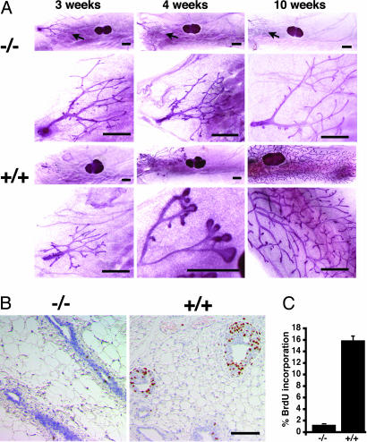
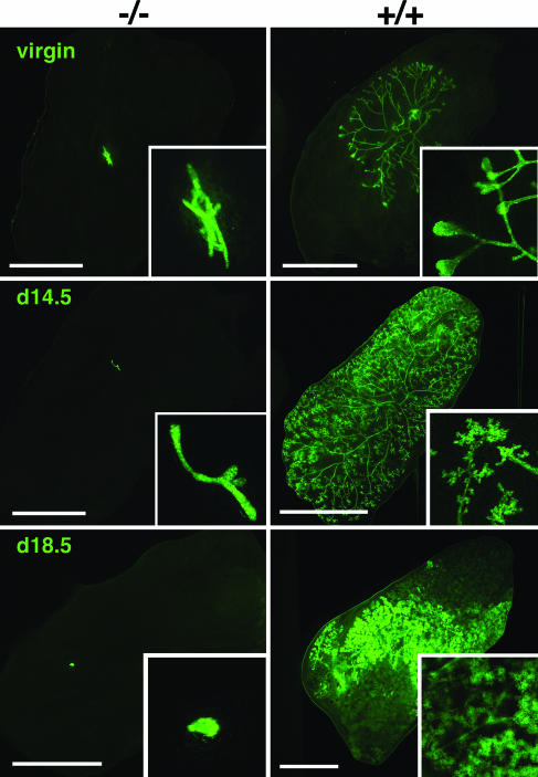
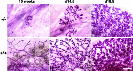
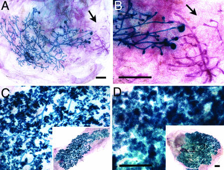
Estradiol is a major regulator of postnatal mammary gland development and thought to exert its effects through estrogen receptor alpha (ERalpha) expressed in the mammary gland stroma and epithelium. Previous studies, however, were confounded by the use of an ERalpha mutant strain that retains some of the protein with transactivation activity. Here, we use an ERalpha-/- mouse strain in which no ERalpha transcript can be detected to analyze mammary gland development in the complete absence of ERalpha signaling. The ERalpha-/- females show no development beyond a rudimentary ductal system. By grafting ERalpha-/- epithelium or stroma in combination with ERalpha WT stroma or epithelium, we show that the primary target for estradiol is the mammary epithelium, whereas a direct response of the mammary stroma is not required for mammary gland development to proceed normally. Mammary glands reconstituted with ERalpha-/- mammary epithelium exposed to pregnancy hormones show increased transcription of milk protein genes, indicating that ERalpha signaling is not an absolute requirement for a transcriptional response to pregnancy hormones. When ERalpha-/- mammary epithelial cells are in close vicinity to ERalpha WT cells, they proliferate and contribute to all aspects of mammary gland development, indicating that estradiol, like progesterone, orchestrates proliferation and morphogenesis by a paracrine mechanism, affecting nearby cells in the mammary epithelium.


Conflict of interest statement

Conflict of interest statement: No conflicts declared.

Figures

Fig. 1.
Fig. 1.
Mammary gland development in ERα−/− mice. (A) Whole-mounted mammary glands of mutant and WT females at different developmental stages. First and third rows show inguinal glands with lymph node. Second and fourth rows show higher magnification of the ductal tree. (Scale bars: 1 mm.) (B) Histological sections of mammary glands stained with an anti-BrdUrd antibody. (C) The percentage of BrdUrd-positive MECs is plotted in the bar graph. A total of 2,000–3,000 cells were counted in three different sections from different mice.
Fig. 2.
Fig. 2.
The role of ERα in the mammary epithelium as shown by transplantation of epithelium. Fluorescent images of mammary glands from recipients at different developmental stages are shown. Preparations were derived from virgin (Top), day 14.5 (Middle), or day 18.5 (Bottom) pregnant recipients engrafted with ERα−/− (Left) or WT (Right) epithelium. (Scale bars: 5 mm.) (Insets) Higher (×5) magnifications are shown.
Fig. 3.
Fig. 3.
Development of mammary epithelium expressing a truncated ERα as shown by transplantation of mammary epithelium from incomplete ERα−/− mice. Whole-mount preparations of mammary glands from WT recipients are shown. Preparations were derived from virgin (Left), day 14.5 (Center), or day 18.5 (Right) pregnant recipients engrafted with incomplete ERα−/− (Upper) or WT (Lower) epithelium. (Scale bar: 0.5 mm.)
Fig. 4.
Fig. 4.
The role of ERα in the mammary stroma. Whole-mount preparations of ERα−/− mammary glands engrafted with WT epithelium are shown in blue. Endogenous epithelium appears red (arrow). Reconstituted mammary glands were removed from the recipients stained with X-Gal before whole mounting. (A and B) Low magnification (A) and a more detailed view (B) of a gland derived from a virgin recipient. (C and D) Days 14.5 (C) and 18.5 (D) of pregnancy. (Scale bars: 1 mm.)
Fig. 5.
Fig. 5.
The role of ERα in differentiation of the mammary epithelium. (A) Hematoxylin- and eosin-stained sections of mammary glands from a recipient engrafted with ERα−/− (Left) or WT (Right) epithelium postpartum. (B) Contralateral mammary glands from virgin (V) mice or mice at different days of pregnancy (days 14.5–18.5) engrafted with ERα−/− GFP+ and ERα+/+ GFP+ epithelia were dissected under UV illumination. RNA was extracted, and cDNA was amplified with primers specific for keratin18, β-casein (Upper), and α-lactalbumin (Lower), and the products were quantified by real-time PCR in triplicate. The relative β-casein and α-lactalbumin expression after normalization for keratin18 is shown. Solid bars indicate the glands engrafted with ERα−/− GFP+ epithelium. Open bars indicate the contralateral glands engrafted with ERα+/+ GFP+ epithelium.
Fig. 6.
Fig. 6.
Rescue of the ERα−/− phenotype in ERα−/− and ERα+/+ chimeric epithelia. (A) Whole-mount preparation of mammary fat pad injected with a mixture of ERα−/− ROSA26 (blue) and ERα+/+ (red) epithelial cells in a 1:10 ratio after X-Gal staining is shown. (Scale bar: 2 mm.) (B and C) Histological section of the same gland counterstained with nuclear fast red. Note that ERα−/− ROSA26 (blue) cells are found in both myoepithelial (arrowhead) and luminal (arrow) cell compartments (B) and are found in TEBs both in the cap cell layer (arrowhead) and among the body cells (arrow) (C).

Similar articles

Cited by

References

    1. Daniel C. W., Silberstein G. B. In: The Mammary Gland. Daniel C. W., editor. New York: Plenum; 1987. pp. 3–31. - PubMed
    1. Brisken C. J. Mammary Gland Biol. Neoplasia. 2002;7:39–48. - PubMed
    1. Nandi S. J. Natl. Cancer Inst. 1958;21:1039–1063. - PubMed
    1. Lyons W. R. Proc. R. Soc. London Ser. B; 1958. pp. 303–325. - PubMed
    1. Lydon J. P., DeMayo F. J., Funk C. R., Mani S. K., Hughes A. R., Montgomery C. A., Jr, Shyamala G., Conneely O. M., O’Malley B. W. Genes Dev. 1995;9:2266–2278. - PubMed

Publication types

MeSH terms

LinkOut - more resources